
Traffic light - Horses
A document that outlines via a traffic light system, the different importance level of antimicrobials for use in horses.
This page will outline antimicrobial guidelines for the following cardiovascular diseases:

Bacterial endocarditis is an uncommon, life-threatening cardiac disorder in horses characterised by bacterial invasion of the endothelial surface of the valves or the wall of the heart, resulting in fibrinous clots or vegetations that impede normal cardiac function. Although bacterial endocarditis can affect horses of all ages, it is predominantly found in animals less than 3 years of age (1).
Cases generally present for evaluation of fever of unknown origin. Clinical signs usually consist of fever with an audible heart murmur (grade 3/6 or louder) on either side of the thorax, with or without tachypnoea, tachycardia, cardiac arrhythmia or signs of congestive heart failure. Vegetative lesions are most commonly found on the mitral valve, followed by the aortic valve. Lesions on the tricuspid valve are less common and pulmonary valve lesions are rare.
Common bacterial isolates are Streptococcus spp., Actinobacillus spp.and Staphylococcus spp. (1, 2), but Serratia marcescens (3), Pseudomonas spp.and E. coli (4) have also been implicated. Cardiac arrhythmias may result from direct extension of the inflammatory lesion into the myocardium or may be secondary to thromboembolic myocardial ischaemia.
Key issues:
Echocardiography can be used to detect thickening of the heart valves. Blood culture is indicated to identify bacteria present and direct antimicrobial therapy. Multiple blood cultures (5 collected at least 12 hours apart) may improve the likelihood of detecting a pathogen. Delaying therapy to allow adequate sample collection is recommended because the identity of the pathogens involved is unpredictable, as is their antibacterial susceptibility, and long durations of therapy are required.
Haematological analysis reveals leukocytosis, with a mature neutrophilia and elevated fibrinogen and Serum Amyloid A (SAA). An electrocardiogram is required if an arrhythmia is detected by auscultation, but this is rare.
Cardiac troponin I has been established as a sensitive and specific marker of myocardial injury (5). Although this is not always present, it could be useful for diagnosis and to monitor the response to treatment.
Broad spectrum antimicrobial therapy is indicated until culture and susceptibility results are available. Resolution of clinical signs, decrease in the size of the lesion on echocardiographic examination and a reduction in leukocytosis, hyperfibrinogenaemia and SAA should determine the duration of therapy, which is likely to be several weeks.
Guarded to poor. Sterilisation of lesions can be difficult to achieve.
Marked chamber enlargement, severe deformation of the valve leaflets, severe valvular regurgitation and signs of congestive heart failure are grave prognostic indicators.
Pericarditis in uncommon in horses and can be secondary to pleuropneumonia or haematogenous spread of bacteria. The pathogens involved reflect these primary diseases. The clinical presentation includes fever, tachycardia and jugular distension. Heart sounds can be heard over a wider than normal area of the thorax, but sounds can be muffled or pericardial friction rubs can be heard. Pericarditis was found more frequently following exposure to Eastern tent caterpillars in the USA but has not been associated with exposure to the processionary caterpillars present in Australia (6).
Key issues:
Echocardiography is required to visualise fluid in the pericardial sac. Thickening of the pericardium with fibrin, coating the visceral and parietal pericardial surfaces, can be evident. An electrocardiogram (ECG) might reveal electrical alternans, where a regularly repeating change in P, QRS and T morphology is present due to regular movement of the heart in the pericardial fluid. Haematology reveals an inflammatory leukogram with elevated fibrinogen and Serum Amyloid A (SAA) concentrations.
Sample collection is often possible when there is moderate to severe accumulation of fluid. Fluid analysis reveals high total protein concentrations and elevated white cell counts with a predominance of neutrophils. Culture and susceptibility testing should be pursued in every case, as the identity of the pathogen is unpredictable and may not be consistent with pathogens cultured from the pleural space or tracheal washes.
Drainage of the pericardial fluid under ultrasonographic guidance is critical in cases where fluid accumulation is moderate or severe. Collection of fluid for bacterial culture and susceptibility testing and cytological examination should be pursued whenever possible.
Broad spectrum antimicrobial therapy is indicated until culture and susceptibility results are available and can be guided by cytological findings. Non-steroidal anti-inflammatory drugs are an important component of medical treatment of pericarditis (flunixin meglumine 1.1 mg/kg IV q 24 h or 0.5 mg/kg IV q 12 h).
Monitoring of pericardial thickening and cardiac chamber dimensions is advisable as pericardial constriction appears to be an important fatal sequela of equine pericarditis that may develop insidiously, one or possibly up to two years after diagnosis (7).
There are no published studies on which to base the duration of therapy, but it is typically 2-3 weeks, or until resolution of the pericardial effusion.
Sprayberry and Slovis (8) followed a small group of young thoroughbred horses and found a reasonably good prognosis, with some attaining a high level of athletic performance, after successful treatment of pericarditis. This study was dominated by cases associated with Eastern tent caterpillars, which are not present in Australia, so the prognosis may vary with other primary causes of disease.
Purpura haemorrhagica (PH) is a non-septic, immune-mediated vasculitis that occurs as a rare complication of strangles (or vaccination against it), due to a type 3 hypersensitivity reaction. Typically, disease develops 2-4 weeks after infection with Streptococcus equi, although it may occur more quickly following vaccination (within a week). Although rare, vaccination with agents containing the SeM protein or avirulent S. equi are associated with an increased risk of PH. Occasionally PH may occur secondary to infection with another infectious organism. It appears to be caused by deposition of immune complexes on blood vessel walls following antigenic stimulation.
Painful pitting oedema results from a necrotising vasculitis. The head, limbs and trunk are most frequently affected with petechiation and ecchymoses of the mucous membranes. Severe oedema can result in exudation from the skin surfaces, and sloughing of the skin may occur. The vasculitis can affect other sites, including the gastrointestinal tract (resulting in colic), lungs (resulting in respiratory distress) and muscles (resulting in infarcts or muscle pain). Fever, tachycardia, tachypnoea, anorexia and depression are also frequently present. Renal dysfunction is common.
Secondary complications are common and can include laminitis and thrombophlebitis. Rarely, serious or even fatal haemorrhage can occur.
In horses with evidence of PH, haematological and plasma biochemical analysis may reveal a leukopaenia or leukocytosis, anaemia, thrombocytopaenia, hyperfibrinogenaemia, hypo- or normoproteinaemia, and increased concentrations of muscle enzymes.
A presumptive diagnosis is generally made based on the history, clinical signs and exclusion of other causes of oedema. Elevated titres of IgA and IgG against S. equi are supportive of a diagnosis. Very high SeM-specific antibody titres (greater than 1:12,800) are strongly suggestive of PH secondary to strangles infection or vaccination. Skin biopsies can confirm leukocytoclastic vasculitis but are rarely performed.
The objective of treatment is to reduce inflammation. Tapering doses of corticosteroids are used – typically dexamethasone, with initial dose of 0.1–0.2 mg/kg IM or IV for 3-5 days and then reducing the dose by half every 3-5 days over 2–4 weeks. Antimicrobials may be necessary where bacterial infection is concurrent or considered likely. Other supportive care such as hydrotherapy, supportive bandaging, IV fluids and non-steroidal anti-inflammatory drugs are usually required.
The case fatality rate is ~10%. Relapse in clinical signs is common if corticosteroids are withdrawn too quickly. Anecdotally, treatment of relapsed disease is more difficult than treatment of horses in their first episode.
Thrombophlebitis is the inflammation of blood vessels in association with thrombus formation. It occurs most commonly due to perivascular injection of irritant substances and can be septic or non-septic.
Septic thrombophlebitis is characterised by heat, swelling, pain on palpation and exudation at the site of venepuncture or around indwelling intravenous catheter sites. Ultrasound is a useful tool to help differentiate between non-septic and septic forms of thrombophlebitis. In non-septic thrombophlebitis there is usually thrombus formation in the vein, without visible fluid pockets or associated perivascular inflammation.
Staphylococcus spp. are the most common organisms cultured from catheter tips in septic thrombophlebitis (11), but E. coli, Streptococcus zooepidemicus and Actinobacillus equuli have all been cultured from cases in Australian horses (12).
The catheter material influences the degree of irritation to the vein and increases the likelihood of thrombosis and sepsis. Polyurethane catheters are less traumatic than those made of polytetrafluorethylene (Teflon) (11). Over-the-wire catheters are generally more flexible than over-the-needle catheters and are thought to cause less injury to the vascular endothelium (13). As a general rule, an over-the-needle Teflon catheter should be replaced after three days, and a polyurethane over-the-needle catheter should be replaced after five days, but replacement should occur earlier if signs of thrombophlebitis develop (13).
Over-the-wire catheters can be left in situ for up to 30 days if good aseptic technique is used during placement. Bacterial endocarditis is a rare, but possible, complication of septic jugular vein thrombophlebitis.
Because of the poor vascularisation that results from thrombosis, septic foci can be difficult to resolve.
Pain, heat and swelling at the site of intravenous injections or of an indwelling catheter, and fever are usually present with septic thrombophlebitis.
Ultrasound can help to establish the extent of the thrombus and the presence of cavitation, which may be indicative of sepsis. Non-septic thrombophlebitis can also occur and can be differentiated clinically by the lack of heat and pain at the site of thrombosis and the absence of cavitation.
Removal of the catheter and culture of the catheter tip are recommended.
Anti- inflammatory drugs used systemically (phenylbutazone 4mg/kg PO q 24 h or 2 mg/kg PO q 12 h or meloxicam 0.6mg/kg PO q 24 h) and hot-packing and topical DMSO can aid resolution of the inflammation.
Broad spectrum antimicrobial therapy is indicated in cases of septic thrombophlebitis until the results of culture and susceptibility testing are available.
Surgical resection of the jugular vein has been described for refractory cases (12).
Most cases resolve with antimicrobial and anti-inflammatory therapy. Some may require surgical resection.
Bacterial endocarditis is a very rare complication and has a poor prognosis.
Vasculitis can involve the arteries, veins or capillaries. Primary disease can occur because of direct damage to the vessel walls by an infectious agent, such as equine arteritis virus. This virus does occur in Australia, but disease associated with it has never been reported, suggesting that the virulence of Australian strains is low. Equine viral arteritis is a notifiable disease in Australia. More commonly in Australia, vasculitis occurs secondary to a bacterial or viral infection or endotoxaemia (14). Vasculitis secondary to infection with Streptococcus equi subsp equi is most common (see purpura haemorrhagica).
Vasculitis can also occur secondary to infection with equine herpesvirus 1 (EHV-1) (15)and, rarely, equine herpesvirus 4 (EHV-4), where there is also an immune-mediated pathogenesis. EHV-1 infection increases the procoagulant activity of peripheral blood mononuclear cell tissue factor (TF). The increase occurs during the acute infection and is most marked at the onset and end of viraemia. Tissue factor is an activator of the coagulation cascade during viral infection.
Horses are usually febrile. The presence of painful, pitting oedema is consistent with the diagnosis. Haematological analysis is useful to determine white cell counts and neutrophil counts. When secondary to endotoxaemia, neutropaenia is common. When secondary to bacterial or viral infection, leucocytosis and neutrophilia are common.
Oedema due to low oncotic pressure should not be painful on palpation. In addition, normal serum albumin and protein, in association with a normal hydration status, rule out decreased oncotic pressure as cause of distal limb oedema.
Diagnosis of the primary disease should be attempted but may not be possible for viral aetiologies. Horses with endotoxaemic vasculitis are generally very unwell, with leukopaenia and neutropaenia. Plasma fibrinogen and SAA concentrations may be elevated during the initial stages, but this is dependent on the stage of disease. EHV-1 and EHV-4 can be diagnosed by isolation of virus from nasopharyngeal swabs or the buffy coat in blood samples collected into EDTA tubes, or by nucleic acid amplification (PCR testing). A rising serum antibody titre can also be used, but results are usually not known until after completion of the clinical course of disease.
Anti-inflammatory therapy is indicated with flunixin meglumine at 0.25mg/kg IV q 8 h (anti-endotoxic dose) up to 1.1mg/kg q 12 h for full anti-inflammatory effect (0.5 mg/kg IV q 12 h is common).
Antimicrobial drugs are not indicated for treatment of vasculitis unless they may be required to treat the primary disease process (for example, procaine penicillin in the case of Streptococcus equi subsp equi infection – see purpura haemorrhagica; Section 2, Chapter 3).
Corticosteroids are indicated for treatment of immune-mediated diseases like purpura haemorrhagica (dexamethasone 0.1 – 0.2mg/kg IV q 24 h; prednisolone 0.10 - 0.2mg/kg PO q 24 h)
Can be guarded depending on the degree of vascular damage that occurs.
Funding for these guidelines was provided by the Australian Veterinary Association (AVA), Animal Medicines Australia (AMA) and AgriFutures Australia.
These guidelines would not have been possible without the considerable expertise and efforts of the Expert Panel: Associate Professor Laura Hardefeldt, Dr. Leanne Begg, Dr. Stephen Page, Professor Glenn Browning, and Professor Jacqueline Norris. We are also extremely grateful to the additional contributing authors.
The dedicated and skilled work of Project Manager Dr. Kellie Thomas is gratefully acknowledged, as are the contributions of the Project Steering Committee: Dr. Phillip McDonagh, Dr. John Messer, Professor James Gilkerson, and Dr. Melanie Latter. Open access publishing facilitated by The University of Melbourne, as part of the Wiley - The University of Melbourne agreement via the Council of Australian University Librarians.
![]()

A document that outlines via a traffic light system, the different importance level of antimicrobials for use in horses.
The Australian Veterinary Prescribing Guidelines cattle and horse flipbook, detailing antimicrobials for use in cattle and horses.
The equine Australian Veterinary Prescribing Guidelines for antimicrobial use as a pocket guide booklet.
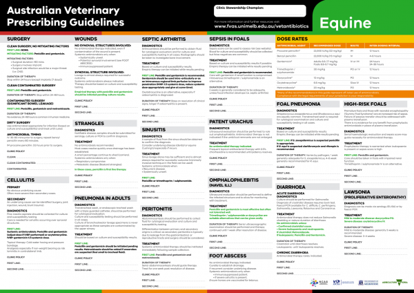
The equine Australian Veterinary Prescribing Guidelines poster. This document that outlines different antimicrobials for use in horses according to different diseases.
Suggest a disease, syndrome or key evidence you think we should include.