
Traffic light - Horses
A document that outlines via a traffic light system, the different importance level of antimicrobials for use in horses.
This page will outline guidelines for antimicrobial use in reference to equine dentistry:
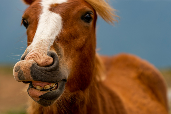
The use of antimicrobials is not indicated in most dental interventions. Even in more advanced cases of dental or sinus disease (see section 5, chapter 2), where bacterial infection is suspected or confirmed, the use of antimicrobials is not consistently necessary. Conservative or tooth-preserving treatment in horses has become an option in recent years, however, our ability to perform endodontics or periodontal treatments is limited by the often-advanced state of dental disease in horses at first presentation to a veterinarian. Dental extraction therefore becomes the most appropriate option in many cases. Preservation of the tooth may be possible when disease is identified in its early stages and antimicrobials play a limited role in our ability to achieve this.
The surface of the equine oral cavity is colonised by a variety of microorganisms, and ~50% of the oral microflora cannot be cultured using conventional techniques (1). Aerobic and anaerobic bacteria have been detected in both healthy and diseased oral cavities (2), with samples from healthy horses showing lower diversity (3). The dominant aerobic genera in the oral cavity of diseased horses were Streptococcus spp. and Actinomyces spp. (4). A shift in the oral bacterial flora towards predominantly anaerobic and Gram-negative bacteria has been described in horses with periapical abscesses, necrotic pulps and periodontal disease (3, 5, 6). Bacterial genera isolated from swab samples of extracted teeth largely corresponded to those identified in blood cultures during and after tooth extraction of these same horses (4).
EOTRH is a disease with an unclear aetiopathogenesis. In horses with EOTRH of the incisors, bacteria from the ‘red complex’, such as Treponema and Tannerella spp., were isolated significantly more frequently than in healthy horses (7). However, antimicrobial therapy has not found to be beneficial in reducing the progression of the disease. At present extraction of the affected teeth is the only valid treatment option available (see details on extraction below).
Transient bacteraemia of oral origin has been described after dental surgical manipulation in humans and dogs, and has also been detected during and after oral extraction in horses (4). The clinical significance of this bacteraemia is unclear, as none of the horses in this study developed complications related to the bacteraemia. Nevertheless, transient bacteraemia may lead to sepsis and other serious diseases of individual organs. Only a few case reports describe infectious and sometimes lethal complications, such as arthritis, endocarditis, meningitis or pneumonia, after tooth removal or sinus surgery in horses (8-10). The bacteria implicated were those that are usually isolated from the equine oral cavity or infected paranasal sinuses.
The severity of the sequelae caused by bacteraemia raises the question of whether horses should be given an antimicrobial drug prophylactically when teeth are extracted (4).The prophylactic administration of antimicrobials is not generally recommended for people undergoing dental treatment. It is known that tooth brushing, normal chewing and the use of dental floss can lead to bacteraemia, and that the risk increases in the presence of dental disease (11). Pre-existing conditions, such as congenital heart disease or artificial heart valves, are an indication for the prophylactic administration of antimicrobials to human dental patients when the apical or gingival tissue is damaged (11). However, antimicrobial drugs cannot prevent the occurrence of bacteraemia and there is controversy in human dentistry about whether antimicrobials can minimise the incidence, severity or duration of bacteraemia, as there is insufficient research to make a determination (12, 13). However, national and international guidelines no longer recommend their use, except under very limited circumstances (14). Ultimately, it cannot be determined whether the risks and costs of antimicrobial administration outweigh any beneficial effect, even preventing severe diseases like endocarditis (12).
Comprehensive evaluation of horses suspected of dental disease is crucial. Examination of the oral cavity with the aid of a mirror is the minimal acceptable practice. A better option to detect subtle changes, such as open pulp cavities, small fistulas, dental cracks or other externally unidentifiable pathologies, is the use of oroscopy. Radiography is a crucial addition to the identification of dental pathology, but its sensitivity and specificity is relatively low. Radiography is particularly poor for the identification of apical infections in the early stages, when antimicrobial therapy may be successful. Computed tomography is the gold standard in the identification of early onset dental disease and should be recommended in cases with suspected disease when radiographic signs are absent.
Randomised controlled trials comparing treatment of horses with dental disease with or without antimicrobials have not been performed. Our clinical experience and reports from case series show that systemic treatment with antimicrobials alone in horses with deep tooth fractures, open pulps or other serious pathological findings is rarely successful. One study reported only 10% of acute apical infection of the caudal maxillary molars with secondary sinusitis respond favourably to systemic antimicrobial therapy. This appeared to be particularly true in young horses with wide open pulp canals and a good blood supply to the common pulp cavity which are far more likely to respond favourably to antimicrobial treatment than older individuals (15). In these cases, a combination of trimethoprim-sulfonamide and metronidazole is recommended for systemic treatment (16). Horses with chronically diseased teeth can usually only be helped by extracting the affected tooth in order to eliminate the cause of the infection and minimise secondary damage (17).
If identified in very early stages, one week of doxycycline (10 mg/kg PO q12h) or trimethoprim-sulfonamide (30 mg/kg PO q12h) may be attempted. Anti-inflammatory therapy is essential.
Through progression of caries, dental fissures or bacteraemia, bacterial contamination of pulpar tissues is possible but the main insult to pulp tissue is the inflammatory response. Once the inflammatory cascade is initiated the confined nature of the pulp chamber leads to a compartment syndrome-like scenario. Increasing intrapulpal pressure compromises vascular supply, leading to pulp necrosis. At this stage, even if bacteria are present, the devitalised tissue no longer permits effective antimicrobial penetration. Therefore, systemic antibiotics have little to no therapeutic value once the pulp is necrotic. Clinical outcomes hinge not on pharmacological intervention, but on timely mechanical management—either by decompression, endodontic therapy (where feasible), or extraction. Relying on antimicrobials alone delays definitive care and contributes unnecessarily to resistance selection.
Endodontic treatment (therapy addressing the inside of the tooth thereby preserving the tooth) has become an option with variable to good results but requires referral to an equine dental specialist. Successful treatment of five out of six cases of apical infection caused by patent infundibula has been described (18). An orthograde endodontic technique applied in 700 cheek teeth reported an 80% success rate in 474 follow up cases (19). More recently a 75% success rate in endodontic treatment and restoration of incisor teeth affected with apical disease was shown (20).
In parasagittal uncomplicated (not exposing pulp cavities) fractures, conservative treatment is favoured. This does not include antimicrobials as no vital tissues are exposed. Monitoring of progression is advised.
In case of complicated fractures with fresh exposure of the pulp cavities, pulp capping is recommended. This also is not an indication for the use of antimicrobials.
Teeth with complicated chronic fractures with evidence of apical disease and sagittal fractures with apical communication are best extracted.
In cases of diastema and associated periodontal disease, local treatment is advisable. Debridement of the affected area, diastema widening, occlusal grooves and specific odontoplasty are all treatments that allow for the management of diastema and periodontal disease. Extraction of teeth with an elevated periodontal disease score (21) may be required. Very rarely are antimicrobials required either locally or systemically. Some recurrent cases of periodontal disease associated with diastema may require local antiseptic application.
Peripheral caries, although including a bacteriological component in their aetiology, are not amenable to be treated with antimicrobials either locally or systemically. The treatment is composed of management of epidemiological factors such as reducing oaten hay in the diet, increasing access to pasture and reducing reliance on concentrate feed and reducing acid (by not feeding silage or acidic water) (22).
Extraction is currently the only viable option for EOTRH affected teeth. The rationale to provide antimicrobials following extraction is debatable in EOTRH cases as infection is present preoperatively and antimicrobial treatment has not been associated with resolution of disease. In the clinical experience of the authors, antimicrobial treatment is not necessary for an undisturbed healing process unless co-morbidities are present. A maximum treatment duration of 5 days is advised.
The administration of antimicrobials to horses undergoing classical uncomplicated oral extractions had no effect on outcome or the level of complications (23).
The preoperative use of antimicrobials for dental surgery is indicated only if secondary diseases such as painful, reactive osteitis (e.g. localised bony swelling with ipsilateral lymphadenitis), purulent fistula tracts, sinusitis or meningitis already exist or if there is a very high risk of them developing. In cases with known immunodeficiency or simultaneous application of high dose glucocorticoids, the use of antimicrobials, and in particular bactericidal antimicrobials, is indicated (24). In horses undergoing minimally invasive transbuccal screw extractions, lower rates of complications have been shown when perioperative antimicrobials (discontinued after surgery) are administered (23).
In cases of mandibular draining tracts, administration of antimicrobials is normally unsuccessful. Temporary resolution may be achieved but considering they mostly occur due to chronically infected dental apices, identification of the causative tooth and extraction or endodontic treatment is the only effective treatment option.
Persistent mandibular draining tracts can also develop following extraction of a tooth. Bone sequestration or remaining teeth fragments in the alveolus are common conditions related to a non-healing, recurrent or persistent fistulisation following extraction of a tooth. Repeat oral inspection and imaging, sometimes requiring CT for proper identification may be required to identify the fragments or sequestrum. Antimicrobial treatment is highly unlikely to result in resolution.
If cellulitis accompanies the draining tract antimicrobials should be administered for 3-5 days.
Table 5.1. Recommendation for perioperative antimicrobials for equine dental disease
Antimicrobial | Dose | Timing | Duration | |
Healthy horse, uncomplicated oral extraction | None OR | |||
Benzyl penicillin OR | 22,000 IU/kg (12 mg/kg) IV | 30 – 60 minutes before the start of surgery | Re-dose every 80 minutes Discontinue after surgery | |
Procaine penicillin | 22,000 IU/kg (22 mg/kg) IM | 3.5 hours before the start of surgery | Single dose only | |
Concurrent disease or when using minimally invasive transbuccal extraction (MTE) | Benzyl penicillin OR | 22,000 IU/kg (12 mg/kg) IV | 30 – 60 minutes before the start of surgery | Re-dose every 80 minutes Discontinue after surgery |
Procaine penicillin | 22,000 IU/kg (22 mg/kg) IM | 3.5 hours before the start of surgery | Single dose only |
In routine cases without co-morbidities horses should be closely monitored post-operatively to detect early signs of complications that could be attributable to bacteraemia or infection, such as fever or tachycardia. The use of serial SAA samples can aid in identification of potential infectious complications. The use of antimicrobials should then be discussed and considered on a case-by-case basis only if problems arise.
Sequestrum formation can follow extraction. In the absence of peripheral signs of wound infection, the administration of antimicrobials is not indicated. Damaged and sequestrating bone requires a long time, sometimes weeks, before demarcating. Once fully sequestrated, removal of the bone fragments will lead to rapid resolution with granulation of the socket and adequate healing. The negative impacts of long duration antimicrobial treatment in these cases far outweighs the potential and unproven benefits.
Prognosis for dental disease resolution is generally good to excellent but is highly reliant on the appropriate diagnostics and removal of the underlying cause. Ongoing alveolar sequestration following extraction, formation of oro-sinus/nasal/cutaneous fistulas, and ongoing sinus discharge are all complex complications of dental disease which require more complex surgical interventions for resolution.
Prognosis for resolution of apical infection is highly dependent on the cause and the chronicity of the infection. In many cases antimicrobials will fail, particularly considering the pulpar tissue present in the teeth is often necrotic and antimicrobial penetration weak to non-existent.
There are many causes of sinusitis in the horse. Determining the precise origin can be challenging but crucial for a successful outcome.
Sinusitis can be primary or secondary. Primary sinusitis is caused by infection with bacteria, viruses or fungi without a predisposition or previous illness. In secondary sinusitis there is a clear underlying cause/pathology that will maintain the infection. Secondary sinusitis occurs, for example, as a result of dental disease, paranasal cysts, trauma, fistulas or neoplasia. The diagnosis, and especially the differentiation between a primary and secondary disease of the paranasal sinuses is not always straight forward but crucial to effectively resolve the infection. Extensive and repeated clinical (oral exam, trans-nasal endoscopy) and imaging diagnostics (radiographs, CT) may be required to pin-point an underlying cause.
Oral bacteria can penetrate into the sinus through the, sometimes very thin, alveolar bone and into the maxillary sinuses. Deep periodontal pockets, fractured teeth, open pulps, tooth root abscesses of various origin are some of the more common dental aetiologies.
A shift in the oral bacterial flora towards predominantly anaerobic and Gram-negative bacteria has been described in horses with periapical abscesses, necrotic pulps and periodontal disease (6, 25). However, research using advanced bacteriology techniques could not differentiate between primary and secondary sinusitis based on culture results (26).
In other secondary sinusitis cases, the inciting cause may not be infectious in origin. Sinus cysts are space occupying structures that are not infectious yet cause recurrent irritation of the sinus that results in chronic discharge. Frontal or maxillary fractures can result in presence of small bone sequestrate that act as foreign bodies and also result in discharge, as do neoplastic processes. Treatment should therefore be focused primarily on removing the underlying cause identified.
Clinical examination is generally not very rewarding in differentiating primary from secondary sinusitis. Elevated temperature, nasal discharge – usually purulent, foetid odour from the nostril, dull percussion of the sinuses are often present for both. Facial swelling over the sinuses may also be present, depending on the underlying cause. On initial presentation, a thorough oral examination should be performed to determine if a clear dental cause of the sinusitis is present (deep periodontal pocket, open pulp(s), fractured tooth, draining tract etc).
Radiology of the sinuses and adjacent teeth maxillary 08 to 11 are required to investigate ongoing sinusitis. This may need to be done after flushing of the sinuses to remove accumulated pus to ensure that extensive fluid lines do not complicate diagnoses of subtle dental causes. Widening of the periodontal ligament space, blunting of the tooth roots, sclerosis of the alveolar bone and a ”halo” around one or more tooth roots are some of the indicators of dental disease. Sensitivity of radiographs for identifying dental disease remains around 75% (27). However, reviews of sinus disease highlight that the secondary causes of sinusitis are not identified in about half the cases on first radiographic examination (28). Apart from the ability to identify dental causes of sinusitis, radiographs are helpful in diagnosing the presence of masses, cysts or simply the accumulation of inspissated pus (most often in the ventral conchal sinus) within the sinus complex.
Computed Tomography is regarded as the gold standard for diagnosis of sinus disease. The sensitivity and specificity of the CT surpasses that of traditional radiography with scans having shown 97% sensitivity (29). Considering the extensive 3D anatomy of the sinus complex, clients should be offered the option to have a CT performed, particularly in cases of chronic or persistent sinusitis.
Scintigraphy has shown a good sensitivity to differentiate between dental and non-dental related sinusitis (30) but CT is superior as scintigraphy remains relatively nonspecific in terms of other aetiological causes of sinusitis.
Ultrasound has limited value in the workup of sinusitis. Unless secondary sinusitis is suspected to be caused by facial trauma, in which superficial bone sequestration is involved. Ultrasound is useful for rapid identification of a sequestrum but again CT is superior. Cases of suture periostitis can also be the origin of secondary sinusitis and identification of sequestra by means of ultrasound is easily achieved in those cases (31)jeh.
Oral endoscopy (oroscopy) has become a critical tool in the investigation of the oral cavity. Dental pathologies extending to the occlusal surface can most clearly be visualised with oroscopy. Detailed examination should focus on occlusal surface of pulp cavities, possibly identifying non vital exposed pulp (look for gas bubbles forming at the surface, use probes to identify). Particular attention should be given to the gingival margins as draining tracts can be present along affected teeth.
Trans-Nasal endoscopy will allow identification of drainage from the sinonasal angle. Nasal discharge is often mistaken for sinusitis, however there are many other origins of nasal discharge like rhinitis, nasal foreign bodies, 06-08 apical infections and others. Before investigating sinusitis cases, it is recommended to confirm the discharge is actually originating from the sinus.
Sinoscopy is best performed through a frontal approach with removal of the bulla of the maxillary septum, as this will provide a full overview of the sinus and may aid in diagnostic as well as therapeutic approach. If available, small diameter flexible endoscopes allow for exploration and intervention which can be done through a transnasal approach with relatively successful outcomes (32).
Acquisition of a sinus sample, via trephination with 14G needle, for culture and susceptibility may be useful in some cases. The results of the C&S will not differentiate between primary and secondary causes. Anaerobic culture should be included but is not always available.
A standard process for evaluating sinusitis cases is useful and is provided here (Figure 5.2).

Figure 5.2. Process for investigating equine cases with suspected sinusitis.
Initial management of non-specific sinusitis may include NSAIDS, mucolytics and adequate drainage (feed the horse on the ground, low head carriage exercise) for 7 days to assess response (Figure 5.1).
Second-line treatment may include a 5-day course of antimicrobial therapy (typically using trimethoprim-sulfonamide or doxycycline), with or without sinus lavage. However, this empirical approach is only justified if a thorough investigation, including additional or repeated examinations, has failed to identify an underlying or secondary cause (e.g. dental pathology, cysts, neoplasia, foreign body). A lack of clinical response to this therapy, or recurrence of sinusitis following withdrawal of antimicrobials, should prompt a more comprehensive diagnostic work-up (see above).
If a tooth/teeth are found to be the cause of the sinusitis, exodontia is required to remove the source of infection. If another secondary cause is identified, removal of the cause (cyst, sequestrum, fungal plaques, inspissated pus etc) is required to achieve resolution. Discontinue antimicrobials until treatment for underlying cause is undertaken.
Prolonged antimicrobial therapy for sinusitis has been associated with development of inspissated pus or fungal overgrowth within the sinus complex. In these cases, further antimicrobial treatment is contraindicated.
If diagnostics do not identify any underlying causes, initial treatment with antimicrobials may be justified. In case of re-occurrence of disease, continuation of antimicrobial therapy is not advised and repeated and/or more advanced investigations should be pursued.
Prognosis for sinus disease resolution is generally good but is highly dependent on the aetiology and relies on the appropriate diagnostics and removal of the underlying cause. Ongoing alveolar sequestration following extraction, formation of oro-sinus/nasal/cutaneous fistulas, ongoing sinus discharge etc. are all complex complications of dental and sinus disease, yet none of them will resolve with antimicrobial therapy alone. More complex surgical interventions are usually required. Nasal discharge resolution is not different between cases having received antimicrobials or not, following dental extraction (33).
A document that outlines via a traffic light system, the different importance level of antimicrobials for use in horses.
The Australian Veterinary Prescribing Guidelines cattle and horse flipbook, detailing antimicrobials for use in cattle and horses.
The equine Australian Veterinary Prescribing Guidelines for antimicrobial use as a pocket guide booklet.
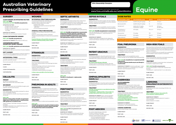
The equine Australian Veterinary Prescribing Guidelines poster. This document that outlines different antimicrobials for use in horses according to different diseases.
Funding for these guidelines was provided by the Australian Veterinary Association (AVA), Animal Medicines Australia (AMA) and AgriFutures Australia.
These guidelines would not have been possible without the considerable expertise and efforts of the Expert Panel: Associate Professor Laura Hardefeldt, Dr. Leanne Begg, Dr. Stephen Page, Professor Glenn Browning, and Professor Jacqueline Norris. We are also extremely grateful to the additional contributing authors.
The dedicated and skilled work of Project Manager Dr. Kellie Thomas is gratefully acknowledged, as are the contributions of the Project Steering Committee: Dr. Phillip McDonagh, Dr. John Messer, Professor James Gilkerson, and Dr. Melanie Latter. Open access publishing facilitated by The University of Melbourne, as part of the Wiley - The University of Melbourne agreement via the Council of Australian University Librarians.

Suggest a disease, syndrome or key evidence you think we should include.