
Traffic light - Horses
A document that outlines via a traffic light system, the different importance level of antimicrobials for use in horses.
Management of retained foetal membranes in the mare requires prompt veterinary intervention, as a delay in treatment leads to an increased risk of potentially fatal sequelae, including toxic metritis and laminitis. Acute metritis can develop, with autolysis of the retained allantochorion or microvilli within the endometrial crypts providing a nidus for infection.The environment becomes ideal for bacterial invasion and replication, leading to inflammation and intrauterine fluid accumulation. Toxins from the bacteria may be absorbed systemically, leading to endotoxaemia, septicaemia and subsequent laminitis (93).
In the mare, the foetal membranes (the foetal portion of the placenta) include the allantochorion, amnion and umbilical cord. These membranes are normally passed during the third stage of parturition, occasionally accompanied by signs of transient abdominal discomfort because of ongoing uterine contractions. Retained foetal membranes occur when there is complete or partial failure of the chorioallantois to separate from the endometrium (93). The equine placenta is diffuse, microcotyledonary and epitheliochorial. Microcotyledons with vascular chorionic villi interdigitate with endometrial crypts to allow increased surface area for nutrient exchange (94). Following parturition, the umbilical cord ruptures, leading to collapse of the umbilical vasculature and smaller vessels throughout the foetal membranes, allowing shrinkage and detachment of the microvilli from the endometrial crypts. Endogenous oxytocin release stimulates rhythmic contractions of the uterine myometrium to expel the free foetal membranes from the uterus (95). In the normal mare, the horns of the allantochorion invaginate as they are released and pass through the ruptured cervical star. This is facilitated by the dependent weight of the membranes (amnion and umbilical cord remnants) hanging externally from the vulva. The membranes are expelled intact, with the allantoic surface outermost.
Foetal membranes are normally expelled within the first three hours post-partum, and membranes retained longer than this are considered ‘retained foetal membranes’ (94, 96). The overall incidence of retained foetal membranes is low in light breeds (2 - 11%) but can be much higher in heavy draught breeds, with the highest prevalence reported in Friesian mares (up to 54%) (97, 98, 99). Although mares may retain foetal membranes following a normal gestation and foaling, several risk factors are associated with a higher incidence of retained foetal membranes, including mare age, breed, a history of retained foetal membranes in a previous pregnancy, and peripartum complications, such as Caesarean delivery, abortion, dystocia, placentitis, prolonged gestation and hydropic conditions (93, 94, 96, 97).
Reaching a diagnosis that a mare has retained foetal membranes may be obvious, as portions of the membranes may protrude externally from the vulva. However, mares may present with concerns of incomplete membrane expulsion or unknown expulsion in the case of an unattended foaling. In many cases, a portion of the allantochorion (i.e. the tip of the non-gravid horn) can be retained and this may not be noticed if the foetal membranes have not been closely examined. Some mares with partial retention of the allantochorion can present 24 to 48 hours post-partum with clinical signs of metritis and/or endotoxaemia, including a fetid vulval discharge, dullness, pyrexia, injected mucous membranes, tachycardia and an increased digital pulse, which is associated with the onset of laminitis (93).
If the membranes have already passed or are expelled following presentation, they should be examined thoroughly to determine whether they are complete and whether there is evidence of pathology. The chorioallantois can be laid out in the shape of an F when it is intact, with the ruptured cervical star at the base and each horn forming the arms. The umbilicus should be located at the base of the gravid horn, with any remnants of the amnion attached. Occasionally the membranes may have been damaged, leading to tearing within the chorioallantois. Often the damaged portions can be pieced together by following the vascular patterns (100, 101).Evidence of incomplete foetal membranes or uncertainty about whether they are complete necessitates further examination of the reproductive tract of the mare.
Further diagnostic procedures are valuable in the diagnosis of retained foetal membranes and metritis. Transrectal palpation of the uterus can be used to determine the degree of uterine tone and involution. Both are reduced with metritis or retained foetal membranes. Transrectal ultrasonography can be used to determine the degree of fluid accumulation and echogenicity within the uterus and may reveal tags of retained chorioallantois. Following cleansing of the perineal area, a transvaginal digital examination can help characterise the nature of fluid within the vaginal cavity and the uterine lumen. Retained membranes can be located and their degree of attachment evaluated. When endotoxaemia is suspected, blood samples should be collected for haematology and plasma biochemical analysis.
Culture of the uterine environment, discharge or uterine effluent is not performed routinely in mares with retained foetal membranes because of the significant level of contamination of the reproductive tract. In cases of ongoing metritis or secondary endometritis, endometrial culture and cytological examination may help determine the need for ongoing antimicrobial therapy.
The objective when managing retained foetal membranes is to attain complete expulsion or removal of the foetal membranes, while avoiding trauma to the entire reproductive tract, minimising excessive force that could lead to uterine horn eversion/uterine prolapse and preventing metritis and endotoxaemia, which can have fatal consequences. There does not appear to be any agreement on or evidence of a ‘best’ method of treatment for retained foetal membranes, nor the timing of when removal should be attempted (102).The method selected for removal of retained foetal membranes is dependent on effectiveness, safety, cost, convenience and the experience of the veterinarian.
The most common method to aid in foetal membrane removal is administration of oxytocin in the early post-partum period. For healthy mares presented within 12 h of routine foaling, conservative therapy with oxytocinmay be all that is necessary. Mares are highly sensitive to the effects of oxytocin in the immediate post-partum period – 10 IU given intravenously or intramuscularly may be all that is necessary for membrane expulsion. The portion of the membranes external to the vulva (if there are any) should be tied in a knot above the hock to ensure they provide constant traction without being trampled or pulled on by the mare. Repeat doses of oxytocin can be considered every two hours for the first 6 h after foaling or until placental expulsion has occurred. Higher doses of oxytocin can result in myometrial spasm and cramping, or signs of colic. Oxytocin can also be administered as a constant rate intravenous infusion (50 - 60 IU oxytocin in 1 L of saline administered slowly over 30 - 60 min) (99) to reduce spasm and signs of colic. In some cases, especially Friesian mares with lower serum calcium levels post-partum, the addition of calcium-magnesium borogluconate (200 ml of a 23% solution) can aid in membrane expulsion (93, 103). Uterine lavage is often combined with oxytocin administration to hasten membrane expulsion.
When the membranes are intact and firmly attached, the chorioallantois can be distended with dilute betadine solution or 0.9% saline (Burns technique), which often will stimulate release of microcotyledons from the endometrium(104). This technique is atraumatic and does not result in any contamination of the uterus when the membranes are intact, without any autolysis. The uterine distension may need to be maintained for up to 30 minutes before membranes are passed, but expulsion often occurs within minutes of distension.
Large volume uterine lavage can be performed to promote separation of the chorioallantois from the endometrial surface. This can be used when the membranes are no longer intact but the fluid runs between the chorion and the endometrial surface (105). Uterine lavage is further indicated when the membranes have been retained for a length of time, which would predispose the mare to metritis. Large volume lavage is used to remove intrauterine accumulations of fluid, inflammatory products, debris and bacteria. Two to 3 litres of warm saline, lactated Ringer’s solution or 0.05% povidone-iodine are infused into the uterine lumen (87). With the hand forming a cage around the tip of the tube, the efflux is removed without aspiration of the endometrium or any retained foetal membrane tags that may remain. The lavage is repeated until the effluent is clear or light pink in colour. In cases of retained foetal membranes following a Caesarean section, uterine lavage should be performed with caution, as excessive fluid distension of the uterus could cause leakage along the suture line (95).
Manual removal of foetal membranes is commonly used, but opinions vary about its use. Techniques that have been described for manual foetal membrane removal include grasping the externalized free portion of the membranes and applying controlled traction (96, 106), placing a hand between the endometrium and chorion to separate the attached membranes in a controlled motion (107), twisting of the allantochorionic membrane into a tight cord (108), and placing a wooden ring between the chorion and endometrium and advancing the ring to separate the membranes from the endometrium (96). The degree of membrane attachment, as well as the duration of membrane retention, can affect the outcome of manual membrane removal, whichever procedure is used. Potential risks of manual membrane removal are a result of excessive traction, leading to retention of the microvilli, tearing and retention of foetal membrane tags, haemorrhage and uterine horn eversion/uterine prolapse. Factors related to the mare (normal vs high risk pregnancy) and the veterinarian (experience, access to the mare) also influence the decision to attempt manual membrane removal.
Another technique, using catheterisation of an exposed umbilical vessel to infuse water, causing distension of membrane vasculature and detachment of the chorioallantois from the endometrium, has been described (109). This procedure leads to stretching of the umbilical vessels, interstitial distension, and subsequent detachment of the microvilli. The result is a rapid but gentle separation of the foetal membranes from the endometrium. The procedure is atraumatic and effective when performed on intact membranes retained for less than 12 h.
Exercise is important to aid in uterine clearance and involution, thus reducing the risk of development of metritis. Paddock turnout is ideal. Hand-walking with frequent uterine lavage and oxytocin therapy can be used when stall confinement is necessary.
Tetanus prophylaxis should be administered if the vaccination status of the mare is unknown. Treatment for endotoxaemia is discussed elsewhere (Section 7).
A survey of equine veterinarians (54% of whom were reproduction specialists) found that the vast variety of treatments for retained foetal membranes reported reflected a lack of specific treatment guidelines and management recommendations. Large volume lavage was the most common treatment used by more than half of the survey respondents. Prophylactic antimicrobials were administered by 42%, with a variety of types, frequency and routes of administration reported (110). The efficacy of antimicrobial use is unknown and intrauterine administration of antimicrobials and antiseptics has been reported to irritate the endometrium and decrease the phagocytic activity of uterine neutrophils (111).
Prophylactic, broad-spectrum systemic antimicrobial therapy should be initiated in mares with suspect metritis or when foetal membranes have been retained for longer than 12 h post-partum. In uncomplicated cases, systemic antimicrobials are continued until uterine lavage is no longer necessary or uterine involution is evident. Intrauterine antimicrobials are not recommended in the immediate post-partum period, with clearing the uterus of fluid and debris using lavage and oxytocin being the preferred option.
Or
The prognosis for survival and future fertility is good in mares that do not develop metritis or endotoxaemia, when resolution of retained foetal membranes is effective and efficient (106, 112). Endometrial culture and cytological examination are recommended on the subsequent breeding cycle to determine if the uterine environment is suitable for breeding. A uterine biopsy maybe useful to evaluate fibrosis if membrane retention has occurred previously. Additionally, mares that have suffered a difficult foaling or dystocia should have a digital cervical evaluation in dioestrus to rule-out cervical abnormalities related to trauma.
The prognosis varies from guarded to moderate in mares that develop metritis, endotoxaemia and subsequent laminitis. The prognosis is further determined by the severity of the metritis, sepsis and endotoxin-induced laminitis, a prolonged disease process and the response to therapy.
A document that outlines via a traffic light system, the different importance level of antimicrobials for use in horses.

The Australian Veterinary Prescribing Guidelines cattle and horse flipbook, detailing antimicrobials for use in cattle and horses.
The equine Australian Veterinary Prescribing Guidelines for antimicrobial use as a pocket guide booklet.
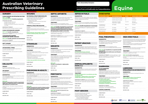
The equine Australian Veterinary Prescribing Guidelines poster. This document that outlines different antimicrobials for use in horses according to different diseases.
Suggest a disease, syndrome or key evidence you think we should include.