
Traffic light - Horses
A document that outlines via a traffic light system, the different importance level of antimicrobials for use in horses.
This causes proliferation of intestinal crypt cells, resulting in a severe protein-losing enteropathy due to thickened walls of the small intestine and, rarely, the large intestine. Lesions are most commonly seen in the ileum, near the ileo-caecal junction. Inflammation is not a characteristic feature but can occur late in the progression of the disease.
The disease occurs worldwide. Affected horses are usually between 4 and 7 months of age. Transmission is faecal-oral. Research suggests that 1 g of faeces from an infected horse would be sufficient to deliver an infective dose to a foal, explaining the exposure rates of up to 100% on affected properties. Foals shed the pathogen for 10-27 days. The bacteria can survive in the environment for at least 1-2 weeks at 5-15 °C (28).
L. intracellularis infects many animal species, including pigs, rabbits, foxes, deer and non-human primates. Rodents appear to be a suitable reservoir host. Properties that have previously housed pigs were initially thought to have been associated with outbreaks in horses, but most cases cannot be linked to exposure to pig faeces and the genomics of pig and equine isolates differ greatly. Year-to-year variation in occurrence on a farm can be expected due to changes in climatic conditions.
Common clinical signs include peripheral oedema (ventrum, penile sheath, throat latch and distal limbs), fever, weight loss, diarrhoea, lethargy and colic. Early clinical signs can be vague and include mild depression, reduced appetite and fever. Diarrhoea can vary from ‘cow pat’ to watery, while some foals have normal faeces. Haematology and serum biochemistry reveal severe hypoproteinaemia due to hypoalbuminaemia. All other blood and serum abnormalities are non-specific and variable. Thickened small intestinal walls can be visualised by transabdominal ultrasonographic examination in many cases. Normal small intestinal wall thickness is less than 3 mm.
Two tests are available for ante-mortem diagnosis. Quantitative real-time polymerase chain reaction assays (qPCR) for pathogen detection in faeces and serological testing (i.e. ELISA). Both tests are recommended, as they have high analytical specificity, but variable sensitivity. In an outbreak in Germany, 21/40 foals tested positive by qPCR, whereas 40/40 were positive by serology (29). Negative PCR results can be expected if the faecal samples are collected from foals that have been treated with antimicrobials or are late in the course of disease. Negative serological tests can be expected in the early stage of the disease. Different PCR and serological assays can also yield variable results.
Clinical signs, in conjunction with hypoproteinaemia in foals on farms with a history of EPE is often sufficient to make a diagnosis. Following diagnosis of a horse on a farm with multiple weanlings, testing of herd mates is strongly recommended. Measurement of total plasma protein using refractometry is an inexpensive and easy way of monitoring at-risk horses. Animals with total plasma protein less than 50 g/L should be subjected to further testing.
Early treatment is critical to avoid advanced disease that results in marked weight loss and critically low serum protein and the need for intensive therapy.
Antimicrobial therapy with oxytetracycline or doxycycline is most common. Early cases can be treated with oral doxycycline (for 7-10 days), but advanced disease should be treated with intravenous oxytetracycline in case gastrointestinal absorption is reduced.
Supportive care is critical in foals with advanced disease, with plasma transfusions, IV fluids and parenteral or partial parenteral nutrition most frequently necessary. Plasma transfusions often result in frustratingly small increases in total plasma protein, even when administered in large volumes (6-8 L). Hydroxyethyl starches (up to 10 ml/kg/day) can be administered, but high doses are associated with coagulopathy and total protein does not increase, so monitoring is difficult unless colloidal oncotic pressure (COP) can be measured.
Total plasma protein can remain low for long periods following clinical recovery.
The prognosis is excellent with early treatment. Spontaneous recovery has not been reported, so treatment is essential. Overall, 93% of treated foals have been reported to fully recover, although mortality in severely affected cases can be as high as 30%.
A document that outlines via a traffic light system, the different importance level of antimicrobials for use in horses.
The Australian Veterinary Prescribing Guidelines cattle and horse flipbook, detailing antimicrobials for use in cattle and horses.
The equine Australian Veterinary Prescribing Guidelines for antimicrobial use as a pocket guide booklet.
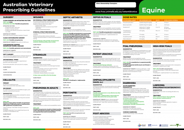
The equine Australian Veterinary Prescribing Guidelines poster. This document that outlines different antimicrobials for use in horses according to different diseases.
Funding for these guidelines was provided by the Australian Veterinary Association (AVA), Animal Medicines Australia (AMA) and AgriFutures Australia.
These guidelines would not have been possible without the considerable expertise and efforts of the Expert Panel: Associate Professor Laura Hardefeldt, Dr. Leanne Begg, Dr. Stephen Page, Professor Glenn Browning, and Professor Jacqueline Norris. We are also extremely grateful to the additional contributing authors.
The dedicated and skilled work of Project Manager Dr. Kellie Thomas is gratefully acknowledged, as are the contributions of the Project Steering Committee: Dr. Phillip McDonagh, Dr. John Messer, Professor James Gilkerson, and Dr. Melanie Latter. Open access publishing facilitated by The University of Melbourne, as part of the Wiley - The University of Melbourne agreement via the Council of Australian University Librarians.
![]()

Suggest a disease, syndrome or key evidence you think we should include.