
Traffic light - Horses
A document that outlines via a traffic light system, the different importance level of antimicrobials for use in horses.
Placentitis is also frequently associated with the delivery of premature or weak foals that may require expensive intensive care if they are to survive (48).
Clinical signs of placentitis may include vulval discharge, premature mammary gland development and other impending signs of parturition (e.g., elongation of the vulva, relaxation of the muscles around the tail head) (49, 50, 51). Vulval discharge is highly variable and only associated with cases of ascending placentitis, and can easily be missed without close and frequent monitoring. Premature mammary gland development is not a pathognomonic sign of placentitis, but rather a non-specific sign of impending pregnancy loss, which can be associated with a variety of other conditions, including twin pregnancy, umbilical cord torsion, poor perfusion and idiopathic/non-specific imminent abortion (51)(figure 1). Enlargement of the mammary gland may also be observed in mares suffering from placental hydrops, pre-pubic tendon rupture and in mares with normal pre-foaling ventral oedema (52). In mares with placentitis, premature mammary development might not be present until the placental inflammation/infection is well advanced.
Four different types of placentitis have been identified, based on their differing morphological lesions and pathogenesis: (1) ascending placentitis; (2) focal mucoid placentitis; (3) diffuse placentitis; and (4) multifocal placentitis (53). The pathogenesis of the different types of equine placentitis are poorly understood. In most cases, infection of the placenta results in placental inflammation and release of prostaglandins, which ultimately leads to abortion or delivery of a premature foal. Foetal infection may also occur, depending on the pathogen involved and the chronicity of the disease process.

Figure 15.3: Common causes of equine pregnancy loss and their associated pathology. Reproduced from: Joan Carrick, Angela Begg, Melinda Stewert, Pat Shearer. AgriFutures Thoroughbred Program.
Several infectious agents are associated with placentitis (Figure 15.3). The primary causes of equine ascending placentitis are Streptococcus equi subspecies zooepidemicus and E. coli, with infection occurring as bacteria ascend through the caudal reproductive tract to the cervical pole region of the chorioallantois (54, 55). Other bacterial agents cultured from cases of placentitis includ, Streptococcus dysgalactiae subspecies equisimilis, Klebsiella pneumoniae, Pseudomonas aeruginosa and nocardioform species. Fungi and viruses can also infect the placenta of mares. However, these pathogens typically cause abortion earlier in gestation (54).Fungi most commonly associated with equine placentitis include Aspergillus spp. and Candida albicans (55). Nocardioform placentitis, a focal mucoid placentitis, is associated with multiple Gram-positive filamentous bacteria. In Kentucky, the most common agents cultured were Crossiella equi, and Amycolatopsis spp. (56). Several cases of focal mucoid placentitis have been reported in Australia (57), but the causative agents may differ from those seen elsewhere. Leptospira spp. are notable causes of placentitis and funisitis in the mare, but no cases of placentitis or abortion attributable to leptospirosis have been reported in Australia (48). A notable cause of placentitis in Australia is Equine Amnionitis and Foetal Loss (EAFL), caused by ingestion of processionary caterpillars. The pathology alone is not specific for EAFL and diagnosis requires demonstration of a combination of pathological and bacteriological features found at necropsy (58). Recently, Chlamydia psittaci has been identified as an important cause of pregnancy loss, premature birth and neonatal loss in Australia and has also resulted in zoonotic disease after contact with infected horses or their tissues (59, 60). While the pathogenesis is not completely understood, the C. psittaci responsible are thought to originate from carrier psittacine birds.
The infection/inflammatory response in placentitis contributes significantly to foetal stress and predisposes to premature parturition or abortion (48). Treatment of placentitis should be aimed at prolonging the presence of the foetus in utero long enough to allow precocious foetal maturation and improved foal survival rates. To achieve a live foal, a prompt diagnosis and effective treatment need to be initiated. Therapy is often necessary before clinical signs are observed to achieve a positive outcome.
Diagnosis of placentitis is often based on a combination of clinical signs and ultrasonographic findings. Unfortunately, clinical signs are often only present in more advanced stages of placentitis. Ultrasonographic examination may be used for both diagnosis and to monitor mares at risk of pregnancy loss. Mares with high-risk pregnancies can be classified into 3 groups: 1) mares with a previous history of abnormal pregnancy, premature delivery or delivery of a septic foal; 2) mares showing clinical signs of an abnormal pregnancy – premature lactation, vulvar discharge, perineal relaxation; and 3) mares with a systemic illness that can affect the foetus, including colic, prolonged surgery, severe lameness, chronic laminitis or systemic infection (48). A complete physical examination should be performed on all mares presenting with clinical signs or for monitoring. Close inspection of the perineal region, the labia and the udder is critical. Any vulval discharge, premature perineal relaxation or precocious mammary development is significant and warrants further examination (49).
Transrectal ultrasonographic examination of the caudal placental pole is commonly used to diagnose ascending placentitis. The correct technique is critical to gain relevant diagnostic information. The clinician should position the transducer with a slight off-midline alignment, with the cervix and ventral aspect of the uterus and placenta at the caudal pole. A ventral branch of the uterine artery/cranial vaginal artery is imaged to ensure correct placement. Three measurements are usually taken of the combined thickness of uterus and placenta (CTUP) and the values can be compared with reported normal ranges (61) (Table 2). Increased CTUP and areas of placental separation are suggestive of ascending placentitis. Improper use of the technique can yield false positive results and should be considered when a clear image is not obtained. Foetal fluid consistency can also be assessed using transrectal ultrasonography. Increased echogenicity can be related to foetal movement, but is also correlated with foetal stress.
Table 15.3. Upper limits for the combined thickness of the uterus and placenta (CTUP) by transrectal ultrasonography during late gestation
Day of gestation | Normal CTUP (mm) |
151 - 270 | < 7 |
271 - 300 | < 8 |
301 - 330 | < 10 |
331 + | < 12 |
(Adapted from Renaudin CD, Troedsson MHT, Gillis CL, King VL, Bodena A. Ultrasonographic evaluation of the equine placenta by transrectal and transabdominal approach in the normal pregnant mare. Theriogenology. 1997;47 2:559-73. (61).)
Transabdominal ultrasonographic examination of the foetus and placenta is used to assess and monitor foetal and placental health (62, 63). It is particularly useful for diagnosis of focal active, diffuse and multifocal placentitis, such as nocardioform placentitis, infection with C. psittaci and cases of EAFL. However, given that a limited area of the uterus is accurately visualized by transabdominal ultrasonography, the apparent absence of pathology does not exclude the possibility of disease. Areas of placental separation with a hyperechoic exudate between the chorioallantois and the uterus are suggestive of nocardioform placentitis (53). The normal amnion appears as a thin membrane surrounding the foetus. Amnionitis can be indicated by a thickened and irregular amniotic membrane (51). Amnionitis, along with funisitis, occurs at the amniotic cord attachment in cases of EAFL and equine psittacosis(59). Foetal heart rate monitoring and the ultrasonographic character of the foetal fluids can also be evaluated (62). A reduced or increased foetal heart rates (normal heart rate is 80 beats/min during late pregnancy) are associated with pathology and are predictive of a poor pregnancy outcome (51, 62). Increased echogenicity of the foetal fluids is often associated with advanced pathology and may also increase with foetal stress and expulsion of meconium, which results in solid particles floating in the amniotic fluid. It is important to note that, in late gestation, increased echogenicity of the amniotic fluid, compared to earlier stages of gestation, is normal.
Cytology and culture of swabs obtained from the external cervical os in pregnant mares can be useful in diagnosis of cases of ascending placentitis when the cervix is open and discharge is present (50). If there is a vaginal discharge, some of the purulent material can be collected aseptically from the caudal vagina without significantly disrupting the vestibular sphincter seal (48). Samples can be collected using a double-guarded cytobrush or swab via vaginoscopy or manually with a sterile rectal sleeve. The presence of inflammatory cells, bacteria, yeasts and/or fungal hyphae will aid the diagnosis of ascending placentitis. A definitive diagnosis of placentitis cannot be made until the foetal membranes are examined for pathology and microbiological investigations are performed after parturition (55). Evaluation of the foetal membranes is a valuable diagnostic tool for cases of known and unknown placentitis related to abortion or neonatal illness (Pozor, 2016). Following abortion, a diagnostic work-up is recommended to determine the cause of foetal loss. Swabs should be collected from the affected membranes for culture and PCR testing for abortogenic agents (e.g., equine herpesviruses types 1 and 4). In addition, foetal tissues (liver, spleen, kidney, heart, and lungs) and foetal body fluids (thoracic and abdominal fluids) can be used for diagnostic testing (64). Because some of the causative agents are contagious (e.g. equine herpesvirus 1) and/or zoonotic (e.g. C. psittaci), appropriate biosecurity measures should be considered when collecting samples.
Several diagnostic biomarkers can be used to monitor placentitis in the mare. Currently, these markers lack the specificity and sensitivity necessary to accurately diagnosis placentitis, but several are helpful in assessing disease progression and response to treatment. In combination with transrectal and transabdominal ultrasonography, the endocrinological markers, progestagins (65, 66), oestrogens (66) and relaxin (67), and the inflammatory biomarker serum amyloid A (68, 69), along with alpha-fetoprotein (70), could be used as additional diagnostic tools. These tests generally require regular monitoring to assess foeto-placental health in late gestation (53). In cases with placentitis, there is frequently an abnormally elevated maternal concentration of progesterone. This is probably a result of dysregulation of progestagen synthesis in the abnormal placenta. Clinical signs of placentitis and low maternal progesterone are indicative of a very poor prognosis for foetal survival. Low oestrogen concentrations in mid pregnancy can indicate severe foetal compromise and imminent abortion. After 280 days of gestation, interpretation of maternal oestrogen concentrations is limited and not well correlated with foetal survival (48). Because the placenta is the sole source of relaxin, plasma concentrations could be used as a biomarker of placental function, but plasma concentrations vary greatly between breeds. Currently, the lack of a commercially available test impedes the clinical use of relaxin assays in mares (48, 67). Alpha-fetoprotein is present in the foetal fluids of mares during late gestation (70), but it remains to be determined whether this protein is a useful marker for spontaneous cases of equine placentitis.
As stated above, placentitis frequently has no associated clinical signs. A preventative monitoring program has been suggested for high-risk mares (48). In one study, mares with a history of abortion or birth of weak, premature/dysmature or septic foals were selected for monthly transrectal and transabdominal ultrasonographic examination. The incidence of ultrasonographic abnormalities in these mares was over 70%. The clinicians were able to initiate treatment based on the ultrasonographic findings. The monitoring program and treatment resulted in a foaling rate of > 90 %, with only 5% of the foals born requiring intensive veterinary care (48).
Current treatment protocols for mares with placentitis are ill-defined and largely empirical. Many treatment strategies aim to combat infection, reduce inflammation and control myometrial activity (71, 72). Therapeutic options include antimicrobials, anti-inflammatories, and progestins (50, 51). Tocolytic therapy has also been suggested, but has minimal clinical efficacy (54).
Although some antimicrobials can cross the placental barrier, foetal fluid concentrations are consistently low. This results in suppression of the growth of bacteria, rather than their elimination. The priority should be to prescribe antimicrobials that cross the placental barrier at doses that maintain sufficient concentrations (MIC) for a duration of time (53). Antimicrobials that have been shown to cross the placenta include trimethoprim/sulphadiazine (73), penicillin and gentamicin (74). The duration of antimicrobial therapy required once a diagnosis of placentitis is made is debated. Anecdotally, short-term antimicrobial treatment (10 – 15 d) has been advocated to be an effective in treating placentitis and avoiding the selection of resistance associated with prolonged antimicrobial therapy (51). Experimental models of induced placentitis do not support the effectiveness of short-term antimicrobial therapy. Mares with experimentally induced ascending placentitis did not carry foals to term when treatment was discontinued after 2 weeks. However, an apparent increase in survival rates was observed when mares were kept on antimicrobials for a prolonged period of time (72). These mares remained culture positive after parturition, suggesting that antimicrobials administered to mares with experimentally induced placentitis may suppress the growth of bacteria, but may not completely eliminate them.
Administration of antimicrobials for 5 - 10 days periodically throughout pregnancy should be avoided. Pulse administration of antimicrobials is often performed in some parts of the world, even in the absence of a diagnosis of placentitis. The idea of such practices would be that this would treat subclinical/undiagnosed placental infection. This practice should be discouraged because of the lack of proven efficacy, and also because it favours selection of antimicrobial resistance that may become a major threat to the health of horses and humans.
Non-steroidal anti-inflammatory drugs should be administered to mares with evidence of placentitis. Flunixin meglumine is very effective in inhibiting prostaglandin synthesis in response to many different inflammatory stimuli, particularly bacterial toxins. Administration of flunixin meglumine (1.1 mg/kg IV q 12 h) is recommended as an initial treatment (72). Once the infection and inflammatory response are adequately controlled, the dose can be reduced or the drug changed (48). Phenylbutazone (2.2 mg/kg PO q 12 h) or firocoxib (0.1 mg/kg, PO, q 24 h) can be administered as an alternative or following cessation of treatment with flunixin meglumine. Firocoxib has the advantage of being a Cox-2 inhibitor, with reduced risks of the side effects typical of NSAID therapy, and it can be administered long-term with limited risk (75).
There is significant synthesis of cytokines by the placenta in response to infection. Pentoxifylline (8.5 mg/kg PO q 8 h) is effective in reducing endotoxin-induced cytokine synthesis. While there are conflicting reports, it may improve oxygenation of the placenta by improving blood flow (48, 72, 76).
The use of altrenogest in high-risk pregnancies is widespread, but its use is controversial because mares with clinical placentitis already have elevated concentrations of progesterone metabolites (65). Most mares with placentitis receive multimodal therapy, including antimicrobials, anti-inflammatories and anti-cytokine drugs, making it difficult to assess the sole impact of altrenogest administration. However, as administration may be very important in maintaining myometrial quiescence, many clinicians continue to administer altrenogest as part of the treatment protocol. It is recommended that, in addition to antimicrobial and anti-inflammatory drugs, altrenogest (44 mg PO q 12 h) is given for an initial 14 days. If there is an adequate response, the dose is reduced to 44 mg q 24 h and maintained until 330 days of gestation. If there is no response, the dose is increased to 88 mg PO q 12 h for 14 days, and the efficacy of the therapy is then reevaluated (48).
Clenbuterol has been used to inhibit uterine contractility. However, the dose required in the mare to reduce uterine contractility can result in significant adverse side effects, including major alterations in cardiac and skeletal muscle function, so its use is not recommended (77).
Intranasal oxygen (to improve foetal oxygenation), vitamin E (antioxidant) and low-dose aspirin (to improve placental oxygenation (78)) have all been administered to high-risk mares. Oestrogen supplementation has been advocated to treat mares with placentitis, as well as treatment with dexamethasone (50), but there are no data available to indicate the success of these treatments.
Repeat ultrasonographic assessment is useful to assess the response to treatment and guide ongoing therapy. The thickening of the placenta, its degree of folding and roughening, and the cloudiness of the fluid will all improve significantly with successful treatment. Mares with a high-risk pregnancy that fail to have any improvement in these ultrasonographic parameters generally have a worse outcome than mares in which improvement is seen.
For ascending bacterial placentitis:
*Penicillin and gentamicin should be used in combination to achieve broad-spectrum therapy unless culture and antimicrobial sensitivity testing has been performed.
The duration of treatment required to control bacterial proliferation is unknown. Studies show that mares with experimentally induced placentitis remain culture positive at the time of foaling despite ongoing therapy (72).
Treatment protocols should be tailored to the individual animal, with treatment duration based on disease progression. Frequent ultrasonographic examination and monitoring are necessary and recommended.
Ascending infection is associated with mares with poor vulval conformation. It remains best practice to perform a Caslick’s procedure or some other surgical correction, such as perineal body repair or uteropexy, in mares with poor conformation.
For diffuse placentitis (EAFL, Chlamydia psittaci)
EAFL is largely associated with abortion without clinical signs and non-specific findings on ultrasonographic examination. To date, there are no reports supporting antimicrobial treatment.
Chlamydia psittaci placentitis is a post-partum diagnosis. Ultrasonographic changes and/or abortion of other mares in proximity may suggest infection (60). Control of chlamydial infections relies on the use of macrolides, fluoroquinolones or tetracyclines (79). There are no evidence-based reports supporting antimicrobial treatment. Empirical antimicrobial therapy using tetracyclines is suggested when ultrasonographic findings are consistent with potential C. psittaci infection (Carrick personnel communication). Premonitory signs are very rare, and are only seen in 5% of cases (64).
For focal mucoid placentitis (nocardioform placentitis)
To date, there are no evidence-based reports supporting antimicrobial treatment of nocardioform placentitis in mares. Therapeutic management of the disease using traditional treatment modalities has had minimal beneficial effect on the foaling outcome. Prophylactic therapy did not decrease the incidence of disease (80).
Early identification and treatment of mares with placentitis has a fair prognosis for foal viability.
It is important to recognise that mares diagnosed with ascending placentitis are at high risk of “red bag delivery” due to thickening and separation of the chorioallantois from the uterus at the cervical pole. These mares should be closely monitored, with foaling attended.
Foals born from mares affected with any form of placentitis are likely to have complications. Neonatal septicaemia is likely if the foal has been exposed to the causative pathogen in utero and the foal may require intensive medical care following delivery.
The mare may also require further treatment after delivery. Even when there has been extended treatment during pregnancy, it is likely that the pathogen remains in the reproductive tract and this increases the risk of metritis and endometritis.
A document that outlines via a traffic light system, the different importance level of antimicrobials for use in horses.

The Australian Veterinary Prescribing Guidelines cattle and horse flipbook, detailing antimicrobials for use in cattle and horses.
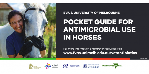
The equine Australian Veterinary Prescribing Guidelines for antimicrobial use as a pocket guide booklet.
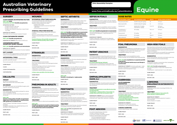
The equine Australian Veterinary Prescribing Guidelines poster. This document that outlines different antimicrobials for use in horses according to different diseases.
Suggest a disease, syndrome or key evidence you think we should include.