
Traffic light - Horses
A document that outlines via a traffic light system, the different importance level of antimicrobials for use in horses.
Click here (https://doi.org//10.1111/avj.70003) to view the guidelines in their entirety.

Corneal ulceration is the result of damage or destruction of parts of the corneal epithelium. This can be initiated by any trauma, exposure injury, a disorder of the cilia, inherited corneal dystrophy, corneal degeneration or calcific band keratopathy. The laterally projecting globes of horses make them particularly susceptible to traumatic ocular injuries. As corneal disease advances, via keratomalacia induced by bacterial, fungal or local host enzymatic destruction, corneal ulceration can involve the corneal stroma, Descemet’s membrane, or may result in a full-thickness corneal perforation. The ocular surface of the horse is constantly exposed to pathogenic bacteria and fungi. Pseudomonas spp., Streptococcus spp. and Staphylococcus spp. are the most common bacterial pathogens involved, while Aspergillus spp. and Fusarium spp. predominate in cases of fungal keratitis. There may be regional variation in the most common microbial pathogens involved (1, 2), and there are multiple reports of emerging antimicrobial and anti-fungal resistance in ocular pathogens in Australia and other parts of the world (1, 3, 4).
A thorough ophthalmic examination performed in a dark stall, with sedation and auriculopalpebral nerve blockade, is necessary to identify a corneal ulcer and assess potential predisposing factors. Fluorescein staining and examination with cobalt-blue light is useful to highlight exposed corneal stroma. A very deep defect down to Descemet’s membrane (a descemetocoele) will not fluoresce. An inability to completely blink may result in an exposure ulcer and therefore the ability to completely blink should be assessed. The adnexa should be inspected for possible distichiasis or ectopic cilia. The surrounding cornea should be assessed for other possible inciting causes and signs of chronicity, such as corneal vascularisation, fibrosis or pigmentation. Initially, corneal/conjunctival swabs should be collected from the affected areas and then immersed in Stuart’s transport medium and submitted for bacterial and fungal culture and susceptibility testing. After collection of a swab for culture, samples should be collected for cytological examination using a Kimura platinum spatula, a cytobrush or the back of a scalpel blade, using a topical anaesthetic specifically formulated for ocular administration, such as proxymetacaine hydrochloride at 5 mg/ml or oxybuprocaine hydrochloride at 0.4%. Fungal hyphae and yeasts have a predilection for Descemet’s membrane but, while detection of them by cytology is confirmatory, failure to observe them by cytological examination does not establish that fungi are not involved. Bacterial, fungal or mixed bacterial and fungal infections can occur in corneal ulcers.
Note: use of a sub-palpebral lavage system will greatly enhance the reliability of drug delivery to the cornea.
No infectious agent observed by cytology:
Cocci observed by cytology:
Rods observed by cytology:
Fungi observed by cytology:
If keratomalacia is observed, use serum eye drops every 2 - 4 h or compounded eye drops containing 3% EDTA every 2 - 4 h (poorly tolerated on corneal ulcers). Serum eye drops are prepared by collecting 6 plain clot vacutainer tubes of blood. They should ideally be incubated at 37°C for 10-15 min to allow the clot to retract and enhance the yield of serum, then centrifuged at 3, 600 x g for 10 min. The serum should then be pipetted or poured off the top and refrigerated. It can be used for 5 days if stored at 4°C.
Atropine should be used in all patients with ulcers, to effect (as observed by mydriasis), for cycloplegia and stabilization of the blood-aqueous humour barrier. The purported association of its use with colic and ileus is dubious (5). Ideally, horses should be housed in a darkened environment if atropine is administered.
Treatment with flunixin at 1.1 mg/kg IV q 12 h, to effect for up to 5 days, or, less ideally, phenylbutazone at 2.2 - 4.4 mg/kg PO q 12-24 h to effect, should be instituted to provide analgesia and reduce the risk of reflex iridocyclitis.
When culture and susceptibility test results are available, and a clinical response to therapy is not apparent, then altering therapy based on the test results is appropriate.
If progression occurs despite appropriate therapy, then globe-sparing surgery is indicated. This may necessitate referral to a veterinary ophthalmologist.
Table 13.1. Antimicrobials used (Corneal ulceration)
Topical Antimicrobial | Use |
Chloramphenicol (10 mg/g) | Gram positives, such as Staphylococcus and Streptococcus spp. |
Polymyxin B (5000 – 10000 IU/g) | Pseudomonas aeruginosa, but significant resistance reported in Australia (1) |
Neomycin (3.5 mg/g) | Pseudomonas aeruginosa, but significant resistance reported in Australia (1) |
Zinc bacitracin (500 IU/g) | Gram positives, such as Staphylococcus and Streptococcus spp. |
Gentamicin (0.3%) | Predominately Gram negatives, also Staphylococcus spp. |
Ofloxacin/ciprofloxacin (0.3%) | Gram positives (such as Staphylococcus and Streptococcus spp.) and Gram negatives (such as Pseudomonas aeruginosa). |
Fair with appropriate management. Despite appropriate management, bacterial colonisation can initiate keratomalacia, which can necessitate rapid surgical intervention. Corneal ulceration is a major cause of vision loss and globe loss in horses.
Fungal keratitis can range from non-ulcerative disease, to rapidly progressive keratomalacia involving all layers of the cornea, to stromal abscessation. The ocular surface of the horse is constantly exposed to pathogenic fungi, so any alteration of host innate defence mechanisms can result in fungal colonisation of the cornea. This colonisation of the cornea has been associated with topical steroid use. The most common species involved include filamentous fungi, such as Aspergillus, Fusarium and Penicillium spp., or yeasts, such as Candida albicans. There are probably regional differences in the fungal species most commonly involved, and there are multiple reports of emerging resistance to anti-fungal agents from around the world (4, 8).
Ulcerative fungal keratitis is a very serious clinical condition and is a leading cause of blindness and globe loss in horses. However, with swift medical, and often surgical, intervention, the globe can be preserved in up to 80% of eyes (9). Fungal keratitis can be slow to resolve medically, with a median healing time of 17 days (range, 12 – 87 days) for Fusarium spp. and 31 days (range, 22 - 63 days) for Aspergillus spp. (4).
See Section 13, Chapter 1.
Note: use of a sub-palpebral lavage system will greatly enhance the reliability of drug delivery to the cornea.
If fungal hyphae or yeasts are observed on cytology:
Topical fluconazole (0.3% solution) has been used q 2-4 h, but it has a more limited spectrum of activity than the other azoles (especially against yeasts).
Silver sulfadiazine (Flamazine) can be used topically q 8-12 h if treatment via subpalpebral lavage is not possible.
If keratomalacia is observed, use serum eye drops every 2 - 4 h or compounded eye drops containing 3% EDTA every 2 - 4 h (poorly tolerated on corneal ulcers). (See Section 13, Chapter 1 for serum eyedrop production and storage).
Atropine and non-steroidal anti-inflammatory use is as recommended for bacterial corneal ulcers (Section 13. Chapter 1.)
When culture and susceptibility results are available and a clinical response to therapy is not apparent, then altering therapy based on the test results is appropriate. Continue treatment until resolution of the keratitis.
If progression occurs, despite appropriate therapy, then globe-sparing surgery is indicated. This may necessitate referral to a veterinary ophthalmologist.
Table 13.2. Antimicrobials used (Fungal keratitis)
Antimicrobial | Use |
Voriconazole at 1% | Good corneal penetration, good historical efficacy against filamentous fungi and yeasts |
Itraconazole at 1% in 30% DMSO | Good corneal penetration, good historical efficacy against filamentous fungi and yeasts |
Ketoconazole at 1% | Good corneal penetration, good historical efficacy against filamentous fungi and yeasts. |
Miconazole at 1% | Good corneal penetration, good historical efficacy against filamentous fungi and yeasts. |
Fluconazole at 0.3% | Yeasts and filamentous fungi but less efficacious than other azoles |
Silver sulfadiazine at 1% in an ointment | Good historical efficacy against filamentous fungi and yeasts. |
With swift and appropriate medical and/or surgical management, vision sparing has been achieved in up to 80% of cases. However, up to 64% of cases require some type of surgical intervention (9).
Stromal abscessation can occur at any depth of the corneal stroma, although it most commonly occurs in the mid to deep corneal stroma. Abscesses form because of micropunctures of the corneal epithelium, with normal epithelial sliding and migration covering the inoculated bacteria and/or fungi, resulting in an abscessed area (11). Environmental factors, such as high wind speed, low temperatures and atmospheric pollutants that disrupt the protective tear film, may contribute to the pathogenesis (12). Horses with stromal abscesses often present with blepharospasm and, initially, a small focal white to yellow opacity in the stroma. An intense iridocyclitis (anterior uveitis) is also often observed.
Covering of the initial epithelial defect with a layer of migrating epithelium can retard the antimicrobial properties of tears and also inhibit access of high concentrations of topical medications to the affected region. Stromal abscesses often require prolonged and aggressive medical and surgical therapy. Deep stromal abscesses are more likely to require surgical intervention than superficial stromal abscesses. Recurrence of abscessation is possible.
A thorough ophthalmic examination should be performed in a dark stall, with sedation and an auriculopalpebral nerve blockade. There should be a careful assessment for iridocyclitis. Fluorescein staining and examination with cobalt-blue light is typically negative, and cytology and culture are often unrewarding.
Note: use of a sub-palpebral lavage system will greatly enhance the reliability of drug delivery to the cornea.
Atropine and non-steroidal anti-inflammatory use is as recommended for bacterial corneal ulcers (Section 13. Chapter 1.)
Signs of improvement include neovascularisation extending throughout the abscessed area, a change in abscess colour to grey-tinged tissue, and control of reflex anterior uveitis.
If progression occurs or appropriate medical therapy does not result in improvement after a week, then surgery is indicated. This may necessitate referral to a veterinary ophthalmologist.
Table 13.3 Antimicrobials used (Stromal abscess)
Antimicrobial | Use |
Chloramphenicol (10 mg/g) | Excellent corneal penetration. Useful against Gram positives, such as Staphylococcus and Streptococcus spp. |
Ofloxacin (0.3%)/ciprofloxacin (0.3%) | Excellent corneal penetration. Broad spectrum, useful against Gram positives (such as Staphylococcus and Streptococcus spp.) and Gram negatives (such as Pseudomonas aeruginosa). |
Voriconazole at 1%: | Good corneal penetration, good historical efficacy against filamentous fungi and yeasts. |
Itraconazole at 1% in 30% DMSO | Good corneal penetration, good historical efficacy against filamentous fungi and yeasts. |
Good for superficial stromal abscesses and fair for deep stromal abscesses. Despite appropriate management, the deep corneal stroma is a difficult location to achieve good drug delivery and extensive corneal vascularisation is needed to achieve complete resolution. Deep stromal abscesses often require surgical intervention.
Equine anterior uveitis is an inflammatory disorder of the equine uveal tract. It can be acute, recurrent or chronic. Uveitis is quite common, occurring in up to 1% of Australian Thoroughbreds (15). The causes of uveitis are often not identified. In cases of uveitis in which a specific cause is identified, the aetiological agent is most often non-infectious, but infectious agents can be involved. A lack of response to symptomatic therapy may necessitate further investigations for infectious causes.
The clinical signs are all due to ocular pain and compromise of the blood-aqueous humour barrier, and include blepharospasm, epiphora, diffuse corneal oedema, aqueous flare, hypopyon and miosis. A particular syndrome of recurrent episodes of uveitis separated by quiescent periods, called equine recurrent uveitis (ERU), is the leading cause of permanent blindness in horses worldwide. Up to 60% of horses diagnosed with ERU do not return to their previous role or perform at a reduced level (16). The development of ERU is complex and multifactorial, but it is probably initiated by a bout of uveitis that alters immune self-recognition via epitope spreading, resulting in autoantigens in various tissues within the uveal tract.
A thorough ophthalmic examination should be performed in a dark stall, with sedation and auriculopalpebral nerve blockade, to diagnose anterior uveitis. The hallmark of active uveitis is aqueous flare, and it may be coupled with corneal oedema, miosis, hypopyon, hyphaema or the presence of fibrin in the anterior chamber. Chronic uveitis may result in posterior synechiae, cataracts, atrophy of the corpora nigra, rubeiosis iridis and retinal detachment. In young animals, primary disease, such as septicaemia and infection with Rhodococcus equi should be considered. If active uveitis does not respond to therapy, then potential infectious causes, such as leptospirosis in endemic areas, should be investigated. Warm, wet environmental conditions support the survival of leptospires in the environment for longer periods, increasing the opportunities for exposure. Paired serum samples can be submitted for serology.
Reduce discomfort with cycloplegics (atropine at 1%), given topically until mydriasis occurs, then continue as needed. The purported association of its use with colic and ileus is dubious (5). Ideally, horses should be housed in a darkened environment if atropine is administered.
Reduce inflammation - topical steroids, such as prednisolone acetate at 1% or dexamethasone HCl at 0.1%, are the most effective, but may potentiate keratomycosis. Topical non-steroidal anti-inflammatory drugs (NSAIDs), such as 0.1% diclofenac sodium, can be used, but these are less effective and can also potentiate keratomycosis. These should be dosed by need, up to 6 times daily, and the frequency of treatment reduced as clinical signs improve. Flunixin meglumine is a potent anti-inflammatory for the eye and can be delivered via intravenous or oral routes. Intravenous administration at 1.1 mg/kg q 12 h is the preferred route. Oral administration of flunixin meglumine is far less effective. Other NSAIDs, such as phenylbutazone, are much less effective in treating the signs of uveitis in horses.
In cases of ERU when inflammation cannot be controlled medically, immune modulation may be warranted. This involves surgical implantation of cyclosporine-impregnated implants into the suprachoroidal space to provide a constant local supply of drug to the uveal tract. This procedure warrants referral to a veterinary ophthalmologist.
Antimicrobials are not used to treat primary non-infectious uveitis, but they are indicated in uveitis that occurs secondary to a specific infectious cause.
Acute uveitis has a good prognosis with appropriate management. Chronic and recurrent uveitis may have a poor prognosis for vision, with the sequelae of cataracts, retinal detachment and glaucoma occurring frequently.
| Fluoroscene | Pupil size | Subpalpebral lavage system? | Antimicrobials | Atropine | Anti-inflammatories | Other therapy | Surgery | |
|---|---|---|---|---|---|---|---|---|
| Ulcers | Positive | Variable | If difficult to medicate | Broad-spectrum q6-8h for ointment, q2h for aqueous. Consider antifungals. | Yes – use to effect for mydriasis | Flunixin IV if hospitalised, Bute PO in field | Serum/EDTA q2-4 for melting ulcers | Yes if: 1. Deep and centrally located 2. Melting 3. Desmetocoele |
Uveitis | Negative | Miosis | If difficult to medicate | None | Yes – q2h until pupil dilated, then as needed | Flunixin IV if hospitalised, Bute PO in the field | No | No |
Stromal abscess | Negative | Variable | If difficult to medicate | Broad-spectrum q6 for ointment, q2 for aquous | Yes – use to effect for mydriasis | Flunixin IV if hospitalised | No | Yes – recommend for all |
Steps for conducting an ocular exam:

A document that outlines via a traffic light system, the different importance level of antimicrobials for use in horses.
The Australian Veterinary Prescribing Guidelines cattle and horse flipbook, detailing antimicrobials for use in cattle and horses.
The equine Australian Veterinary Prescribing Guidelines for antimicrobial use as a pocket guide booklet.
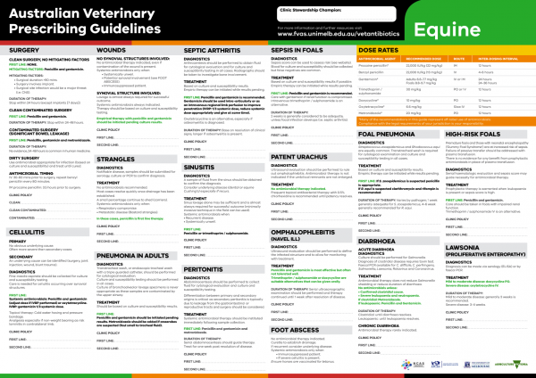
The equine Australian Veterinary Prescribing Guidelines poster. This document that outlines different antimicrobials for use in horses according to different diseases.
Funding for these guidelines was provided by the Australian Veterinary Association (AVA), Animal Medicines Australia (AMA) and AgriFutures Australia.
These guidelines would not have been possible without the considerable expertise and efforts of the Expert Panel: Associate Professor Laura Hardefeldt, Dr. Leanne Begg, Dr. Stephen Page, Professor Glenn Browning, and Professor Jacqueline Norris. We are also extremely grateful to the additional contributing authors.
The dedicated and skilled work of Project Manager Dr. Kellie Thomas is gratefully acknowledged, as are the contributions of the Project Steering Committee: Dr. Phillip McDonagh, Dr. John Messer, Professor James Gilkerson, and Dr. Melanie Latter. Open access publishing facilitated by The University of Melbourne, as part of the Wiley - The University of Melbourne agreement via the Council of Australian University Librarians.
![]()

Suggest a disease, syndrome or key evidence you think we should include.