
Traffic light - Horses
A document that outlines via a traffic light system, the different importance level of antimicrobials for use in horses.
Lesions are usually confined to the proximal half of the small intestine, including all the duodenum and a variable amount of the jejunum, but in severe cases 90% of the small intestine can be involved. Duodenitis/proximal jejunitis is characterised by acute onset of fever, tachycardia, abdominal pain, ileus and the presence of reflux on placement of a nasogastric tube. The severity of pain varies from mild to severe, which manifests in some horses as inappetence, depression and a tendency to recumbency. Clinical signs of endotoxaemia, such as injected mucous membranes, a slow capillary refill time, dehydration, fever and tachycardia are common.
Numerous aetiological agents have been proposed, including clostridial species, mycotoxins and Salmonella spp., but the evidence is weak for all of them. Diet appears to play a role in some horses.
The major differential diagnosis is small intestinal obstruction, but these horses should not be febrile initially and their pain usually becomes more severe and does not completely resolve with gastric decompression, unless the affected viscus ruptures. However, differentiating duodenitis/proximal jejunitis from small intestinal obstructions can be a challenging.
Clinical examination will reveal colic, fever and the presence of large volumes of gastric reflux. Fever may only be present in 25-30% of cases. Mild to moderate colic that resolves following gastric decompression is a hallmark of the disease, with signs of severe colic less common. The colour of the reflux is usually yellow-green to red-brown, and it usually has a fetid smell.
Abdominal ultrasonographical examination reveals a thickened and distended small intestine with no motility (ileus), which can also be palpated on rectal examination.
Abdominocentesis generally reveals peritoneal fluid with an elevated protein (> 35 g/L) and a normal total nucleated cell count (26).
Haematology is not distinctly different from horses with small intestinal strangulating lesions. Serum biochemistry often reveals increased liver enzymes and total bilirubin as a result of compression and occlusion of the bile duct or ascension of disease to the liver via the bile duct.
Therapy is aimed at relieving gastric and intestinal distension, addressing dehydration and electrolyte abnormalities, alleviating pain and promoting gastrointestinal motility.
Regular decompression of the stomach by enabling reflux through an indwelling stomach tube, sometimes as often as every 2 h, is important to reduce pain and ileus and prevent gastric rupture.
Intravenous fluid therapy is required to treat the dehydration from fluid and electrolyte loss through gastric reflux and the inability to drink. Serial monitoring is required to adjust fluid therapy to compensate for ongoing losses through reflux.
Prokinetic therapy is commonly used to combat ileus, with drugs such as bethanechol, phenothiazine, metoclopramide, cisapride and lidocaine all reported, but there is only weak evidence to support its use. A lidocaine infusion (loading dose of 1.3 mg/kg, followed by a constant rate of infusion of 0.05 mg/kg/min), is often used as a prokinetic drug and for pain relief because of its anti-inflammatory properties. Adequate pain relief is critical in combating ileus.
Antimicrobial therapy is controversial. Some advocate for coverage of clostridial species. Penicillin alone or metronidazole are common, but oral metronidazole is ineffective because of the ileus, so penicillin is probably a better choice. Benzyl penicillin IV is a better choice than procaine penicillin IM because of the poor muscle perfusion caused by dehydration and endotoxaemia early in disease. Rectal administration of metronidazole is possible in a refluxing horse, but results in lower bioavailability compared to oral administration so doses of 20mg/kg bid should be used(27).
Treatment of endotoxaemia is covered elsewhere (see Section 7).
Cryotherapy for all four feet is indicated to reduce the risk of laminitis secondary to acute enteritis. Ideally, cryotherapy should be applied for the duration of the developmental phase of laminitis, so horses exhibiting signs of endotoxaemia should be treated when first seen. Continuous application is likely to yield the best results and should be continued until resolution of the primary disease, with some experts continuing for another 24-48 h after resolution of systemic inflammation. Immersion of the limb from the upper metacarpus and metatarsus distally in an ice and water mixture effectively achieves cooling to less than 10 oC, although constant ice replenishment is labour intensive. Commercially available wader-style devices can be modified as an alternative. Although the degree to which the limb must be cooled to prevent laminitis has not been established, current recommendations are for a hoof temperature of 10 oC, although there may be some benefit from even mild cooling (14).
Generally, the prognosis is guarded, depending on the length of time that gastric reflux persists. Laminitis has been reported to occur as a sequela in 30% of cases.
Survival rates of 25-94% have been reported. Case series have been small, so a wide range in survival rates is not surprising.
An anion gap of >15 mEq/L has been associated with 6 fold higher odds of mortality (26).
A document that outlines via a traffic light system, the different importance level of antimicrobials for use in horses.
The Australian Veterinary Prescribing Guidelines cattle and horse flipbook, detailing antimicrobials for use in cattle and horses.
The equine Australian Veterinary Prescribing Guidelines for antimicrobial use as a pocket guide booklet.
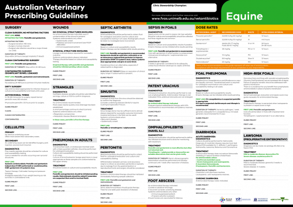
The equine Australian Veterinary Prescribing Guidelines poster. This document that outlines different antimicrobials for use in horses according to different diseases.
Funding for these guidelines was provided by the Australian Veterinary Association (AVA), Animal Medicines Australia (AMA) and AgriFutures Australia.
These guidelines would not have been possible without the considerable expertise and efforts of the Expert Panel: Associate Professor Laura Hardefeldt, Dr. Leanne Begg, Dr. Stephen Page, Professor Glenn Browning, and Professor Jacqueline Norris. We are also extremely grateful to the additional contributing authors.
The dedicated and skilled work of Project Manager Dr. Kellie Thomas is gratefully acknowledged, as are the contributions of the Project Steering Committee: Dr. Phillip McDonagh, Dr. John Messer, Professor James Gilkerson, and Dr. Melanie Latter. Open access publishing facilitated by The University of Melbourne, as part of the Wiley - The University of Melbourne agreement via the Council of Australian University Librarians.

![]()

Suggest a disease, syndrome or key evidence you think we should include.