
Traffic light - Horses
A document that outlines via a traffic light system, the different importance level of antimicrobials for use in horses.
Click here (https://doi.org//10.1111/avj.70003) to view the guidelines in their entirety.

Arytenoid chondritis is a performance limiting upper airway disease most commonly reported in young thoroughbred horses (1). The aetiology is thought to be secondary to trauma to the arytenoid cartilage, with subsequent extension of mucosal bacterial or viral infection into the cartilage. The cartilage infection often progresses, to result in permanent enlargement or deformation of the arytenoid cartilage, requiring arytenoidectomy or resection of a granuloma. Clinical signs include insidious and progressive onset of respiratory noise, exercise intolerance and/or coughing. Bacterial cultures from the infected cartilages and granulomas have revealed mixed infections, with 58% growing Gram-positive bacteria, 54% Gram negative bacteria and 33% anaerobic bacteria (Johnston and Lumsden, 2020). Streptococcus spp. were the most common (32%), followed by members of the Enterobacteriaceae (13%). In this report, bacterial isolates were susceptible to ceftiofur (83%), ampicillin (64%), tetracycline (48%), enrofloxacin (45%), trimethoprim-sulphadiazine (41%) and gentamicin (18%). Susceptibility of bacteria often varies substantially by geographic area, so local data should drive decision making. Ceftiofur and enrofloxacin are high importance antimicrobials and should not be used as first line therapy.
Endoscopy of the larynx is the most widely used diagnostic tool, but ultrasonography of the larynx is useful to determine the extent of the chondropathy.
Culture of the cartilage resected during arytenoidectomy or removal of the granuloma is important, but culture of the surface of these lesions during upper airway endoscopy is not considered useful.
Broad spectrum antimicrobial therapy is indicated. Penetration into the infected cartilage is an issue, as vascularisation is poor. Penicillin and gentamicin are recommended, but oral therapy is often used as longer courses of intramuscular antimicrobials can be poorly tolerated by yearlings. Antimicrobial therapy is usually required for at least five to seven days, when some improvement of the endoscopic appearance is expected. If there is no improvement apparent on repeat endoscopic examination, arytenoidectomy or resection of the granuloma should be considered if there is an impediment to the airway and a future athletic career is anticipated. Culture and susceptibility testing should be performed on the resected infected cartilage and granuloma. The total course of treatment can be weeks, depending on rate of resolution of these lesions.
Anti-inflammatory therapy with phenylbutazone (2 g IV or PO q 24 h for the first five days of treatment) is a useful adjunctive therapy.
Reduction in the lesion size and improvement in arytenoid movement after antimicrobial therapy is encouraging. The prognosis for performance following arytenoidectomy is considered guarded due to the propensity of recovered horses to aspirate.

Epiglottitis can occur with ulceration of the epiglottis and in severe cases can lead to exposure of the epiglottic cartilage (Figure 3.2. Image A). Mucosal lesions most result from trauma and this allows extension of mucosal bacterial and viral infection into the cartilage. Epiglottic entrapment is a cause of exercise intolerance and inspiratory and expiratory noise. The epiglottis becomes enveloped by the aryepiglottic fold (Figure 3.2 Image B).

Figure 3.2: A. Epiglottic entrapment in a horse. B. Epiglottitis in a horse. (Images courtesy of Leanne Begg.)
Both conditions are diagnosed by endoscopy. With epiglottic entrapment, the distinct serrated margins of the epiglottis and the dorsal epiglottic vascular pattern are obscured by a fold of aryepiglottic mucosa. Cartilage involvement is diagnosed when there is thickening and disfigurement of the cartilage of the epiglottis.
Mucosal lesions are best treated with rest and anti-inflammatory drugs. If the epiglottic cartilage is exposed, broad spectrum antimicrobial therapy is indicated.
Epiglottic entrapment requires surgical division of the aryepiglottic fold, which is usually performed standing if it is permanently entrapped but needs to be done under general anaesthesia if it is only intermittently entrapped.
Epiglottitis generally has a good prognosis after antimicrobial therapy unless infection of cartilage leads to disfigurement and compromise of epiglottic function.
Epiglottic entrapment has a good prognosis for future athletic performance following surgery.
Subepiglottic cysts are an uncommon cause of respiratory noise in young horses. They are usually present from birth but are diagnosed as horses begin to exercise, when they may cause exercise intolerance or respiratory noise. Large cysts are diagnosed earlier in foals, as they lead to coughing, dysphagia and food aspiration.
Subepiglottic cysts are diagnosed by endoscopy.
Pharyngeal cysts are removed by surgical resection.
Subepiglottic cysts have a good prognosis following surgical resection.
Pharyngeal lymphoid hyperplasia (PLH) usually occurs in young horses. These follicles are lymphoid tissue that spreads over the larynx. Hyperplasia occurs with lymphoid stimulation. They are usually considered of little significance and not a cause of poor performance.
PLH is diagnosed by endoscopy (Figure 3.3).

Figure 3.3. Pharyngeal lymphoid hyperplasia in a young thoroughbred. (Image courtesy of Leanne Begg.)
No treatment is recommended for pharyngeal lymphoid hyperplasia. Rest and anti-inflammatory drugs may be indicated if it is severe.
Pharyngeal lymphoid hyperplasia has excellent prognosis for future athletic performance.
Fungal infections of the upper respiratory tract are more commonly seen in warm and humid climates, but they do occur rarely in Australia. Aspergillus species are the fungi most commonly isolated from sinonasal mycotic infections in cold climates (1), but three cases of cryptococcal infection have been described in Australia (2-4) and Conidiobolomycocosis is also seen. Clinical signs include dyspnoea, abnormal respiratory noise and decreased airflow through the nares, a unilateral or bilateral nasal discharge that may be malodorous, and bony swelling over the sinuses.
Clinical signs of guttural pouch mycosis include unilateral or bilateral epistaxis, nasal discharge and dysphagia. Epistaxis occurs because of the erosion of the guttural pouch mucosa by the fungal plaque, resulting in haemorrhage from the internal carotid, occipital or maxillary arteries. Severe haemorrhage can result in exsanguination and death (5). Aspergillus species are the fungi most commonly isolated from cases of guttural pouch mycosis (6).
Upper airway endoscopy, including into the guttural pouch, is usually diagnostic for both sinonasal and guttural pouch mycosis. Superficial fungal plaques, as most often seen with Aspergillosis, can be sampled using a cytology brush. For granulomatous masses in the nasopharynx, deep biopsy samples are required. These samples need to be collected with a uterine biopsy instrument as the small endoscopic biopsy instruments are usually too superficial to be diagnostic.
In cases of guttural pouch mycosis, the diagnosis is generally made by endoscopy alone, in conjunction with the clinical history, because of the risk of haemorrhage from disturbance of fungal plaques on arteries.
Serological testing using latex agglutination to identify cryptococcal capsular antigen (LCAT) may be useful if Cryptococcus spp are suspected based on cytological examination.
Where samples can be safely collected, fungal culture should be pursued to confirm the diagnosis and allow for susceptibility testing. This includes cases of sinonasal mycotic infections, which also require imaging studies and are discussed further in Chapter 5.2 Sinusitis (see Section 5 - Dentistry).
Surgical treatment of guttural pouch mycosis is traditionally aimed at obstructing blood flow through at-risk vessels by ligation, balloon-tipped catheter placement or transarterial coil embolism, which also results in inhibition of fungal growth, probably because of the reduction in the availability of oxygen (7). Antifungal medical therapy (systemic or topical) is not required. Laser salpingopharyngostomy into the guttural pouch has been used more recently as an adjunct to treatment of guttural pouch mycosis to alter the guttural pouch environment. This has been shown to specifically alter the oxygen and carbon dioxide concentrations, but not the temperature and humidity, within the guttural pouch (8) and is an alternative option for treatment when the fungal plaque is not associated with haemorrhage. When lesions are located over major arteries then there is always the risk of sudden fatal haemorrhage. If surgical intervention is not available, and the lesions are not over major arteries then repeated topical therapy with nilconazole or nystatin has been used successfully. When lesions affect cranial nerves and dysphagia results, then horses may need prolonged feeding of gruel via a nasogastric tube and possibly treatment for aspiration pneumonia.
Sinonasal mycotic infections caused by Conidiobolomycosis coronatus and Cryptococcus spp are usually successfully treated with 1-3 months of oral fluconazole. Antifungal susceptibility testing is warranted for other organisms, including Aspergillis spp. Not all laboratories offer this service, so it is worth discussing this with laboratories prior to sample submission. Extensive infiltration can be surgically debulked prior to lavage and oral antifungal therapy. One case of cryptococcal rhinitis in Australia was successfully treated with sinonasal bathing with fluconazole under general anaesthesia (4).
Surgical therapy alone is generally successful for guttural pouch mycosis and reduces the risk of exsanguination.
For superficial lesions:
Nystatin or amphotericin B have also been administered via inhalation through a nebuliser twice daily most commonly for 2 weeks. The efficacy and effectiveness of this therapy is unknown as all cases have also been treated with topical therapy (1).
Superficial plaques due to Aspergillus fumigatus should be treated based on results of culture and susceptibility
Extensive cryptococcal granulomas, lesions probably benefit from surgical debulking followed by:
Good for guttural pouch mycosis. Reasonable with prolonged treatment, with reports of one to five months needed to see resolution of fungal plaques when medical therapy is used. Recurrence, sometimes years later, is frequently reported.
Streptococcus equi subspecies equi (S. equi) are beta haemolytic Gram-positive cocci.Considered a pathogen of the upper equine respiratory tract, S. equi is closely related to Streptococcus equi subspecies zooepidemicus (S. zooepidemicus). Unlike S.equi, S. zooepidemicus is considered a commensal organism of equine mucosa and an opportunistic pathogen. The genetic similarity between these two bacterial subspecies has clinical relevance, as it may complicate serological testing and agent identification in many assays.
Stranglesis a highly contagious upper respiratory tract disease of horses. A proportion of affected horses will establish an infection in the guttural pouch and remain persistent carriers of S. equi. Most carriers clear their infection after several weeks, but some remain infectious for months to years. Outbreaks in training yards, agistment facilities and on farms are not uncommon and can be difficult to control. Identification of carriers is challenging, so detection of horses in the early stages is critical, as effective isolation of large groups of animals can be logistically difficult. Unidentified carriers, nose-to-nose contact, fomite transfer on personnel and equipment and in shared water or feeding troughs may perpetuate the outbreak. Strangles is a notifiable disease in some Australian jurisdictions and practitioners are encouraged to inform and seek advice from their local veterinary authorities.
Infection with S. equi in horses generally produces a pyretic episode that precedes clinical signs of upper respiratory tract disease, such as muco-purulent / purulent nasal discharge, pharyngitis and subsequent abscessation of the draining lymph nodes. The severity of the clinical disease may be affected by the age and immune status of the horse. Fever typically occurs 3-14 days after exposure and generally persists until the lymph node abscesses rupture. The substantial pharyngitis results in reluctance to eat and drink. Lymphadenopathy is a typical clinical sign – classically affecting the submandibular and retropharyngeal lymph nodes, although the parotid and cranial cervical lymph nodes are also occasionally involved. Abscesses generally rupture between one and four weeks after infection. Guttural pouch infection and empyema results from abscessation of the retropharyngeal lymph node and subsequent rupture of the abscess into the guttural pouch. Some of these carrier horses develop aggregations of inspissated pus and debris, known as chondroids, which play a role in shielding the bacteria from the immune response and prolonging persistent infection.
Inflammation associated with pharyngitis and formation of lymph node abscesses may cause obstruction of the upper respiratory tract (hence “Strangles”) necessitating temporary tracheotomy.
Although rare, metastatic abscesses can also occur in multiple sites, including the abdomen, and these cases are commonly referred to as “bastard strangles.” There is a clinical impression that the rate of bastard strangles is higher in horses treated with antimicrobials but there is no evidence one way or the other in the literature. Other complications following strangles infection are also well recognised and include purpura haemorrhagica (see Section 2, Chapter 3), and immune mediated myositis (Section 11, Chapter 2). Myocarditis is a less commonly reported sequela of strangles infection.
The overall complication rate increases with the duration and intensity of exposure and may be as high as 20%. Overall case fatality rates can be as high as 8.1% to 9.7% in large farm outbreaks.
Although the clinical signs of strangles are highly indicative, collection of an appropriate sample and identification of the organism is critical, as infection with S. zooepidemicus can mimic the clinical signs of strangles. In the early stages of disease an aspirated sample of pus from an abscessed lymph node provides the ideal, least contaminated sample. Swab samples from burst lymph node abscesses may be of some value if samples are collected carefully to minimise contamination with skin commensals. PCR methods have greater sensitivity for detection of S. equi than traditional culture methods, as have newer molecular methods, such as isothermal assays such as loop-mediated isothermal amplification (LAMP).
In horses with evidence of clinical disease due to S. equi, a complete blood count is non-specific with leukocytosis characterized by a neutrophilia and often hyperfibrinogenaemia. These changes are indicative of a non-specific inflammatory response.
Serial nasopharyngeal swabs and/or guttural pouch washes are often used to detect S. equi following resolution of clinical disease. This sampling is important, as shedding of S. equi after infection may be a source of infection of new horses and thus prolong the outbreak. A minority of horses may continue to shed the organism, maintaining a carrier state via colonization of guttural pouches (with or without chondroid formation). It is important to allow horses to recover from the acute disease before sampling to identify persistent carriers (Figure 3.4).
Several commercial ELISAs are available targeting IgG antibodies against surface proteins of S. equi. These tests have differing sensitivities and specificities, depending upon the type used and stage of disease. The duplex iELISA is useful to manage an outbreak of strangles, as recent infection can be detected by an increase in antibody levels in paired sera taken 10-14 days apart). Other antibody assays, such as the SeM ELISA assay, may detect antibodies directed against SeM of S. zooepidemicus and this cross-reaction leads to reduced specificity of the assay (and false positive results). Microbiological testing of guttural pouch lavages by qPCR or culture can be used to identify sub-clinically infected persistent carriers at the end of an outbreak (Tables 3.1 and 3.2). Sampling for persistent S. equi shedding should commence no sooner than 3-4 weeks after the resolution of clinical signs.
The SeM antibody response can be used to detect horses at risk for development of purpura haemorrhagica, with the recommendation from the USA that horses with strong antibody responses are not vaccinated against S. equi until the antibody levels have subsided.
Fever without clinical signs in a strangles outbreak may be an indication for antimicrobial therapy if there is certainty that other clinical signs have not commenced. For this condition to be met, horses must be able to be kept isolated and body temperature monitored twice daily to ensure that treatment is commenced immediately following development of fever. Early treatment may reduce formation of abscesses and therefore shedding. However, if abscesses have already started to form, antimicrobial therapy is highly unlikely to result in bacteriological cure and will only delay abscess rupture thereby prolonging the clinical course and that of the outbreak. Procaine penicillin is the drug of choice.
Uncomplicated strangles infection can usually be managed using supportive treatments. Lancing abscesses to establish drainage will hasten recovery, but most abscesses burst and drain, either externally or internally, within one to four weeks. Non-steroidal anti-inflammatories can reduce fever and improve appetite until drainage is established. Bathing and lavage of burst abscesses and non-steroidal anti-inflammatory medications generally results in rapid healing. Despite a perceived indication by many practitioners and lay personnel, the use of antimicrobials is not indicated in uncomplicated cases of strangles and may delay recovery, especially when there is lymphadenopathy (abscess formation) and drainage has not been established.
Antimicrobials are indicated in infected horses with respiratory distress where abscess drainage is not possible or does not resolve airway obstruction. The antimicrobial drug of choice is penicillin given the predictable sensitivity of S. equi to this antimicrobial.
Oral antimicrobials are sometimes preferred for logistical reasons and trimethoprim-sulfadiazine (TMS) [30 mg/kg PO q 12 h] has in vitro efficacy against S. equi, however anecdotal reports of clinical efficacy vary. In emergency situations, penicillin should be used as streptococci are highly susceptible.
Guttural pouch empyema may require repeated lavage with isotonic saline or polyionic fluids. Both topical and prolonged systemic penicillin administration (10 days) have been used successfully to treat these cases. Endoscopic basket removal of any chondroids is preferred over surgery, as complications with surgery are common.
Metastatic abscessation is considered a complication of a small proportion of strangles cases and is commonly referred to as “bastard strangles”. Sites of infection include internal abdominal organs, lungs and brain. These cases remain difficult to diagnose and need to be treated on a case-by-case basis. As with all abscesses, drainage is the key to clinical resolution. Considerations influencing antimicrobial selection administration include efficacy against S. equi but also include formulation suitable for long-term administration and ability to penetrate tissue (high lipid solubility). Procedures to assist local drainage are indicated, if practical.
Metastatic strangles: penicillin 22,000 IU/kg IM q 12 h (if tolerated long term) or trimethoprim-sulphadiazine at 30 mg/kg PO q 12 h – treatment should continue until the abscesses are no longer visible on ultrasound (which may be several weeks to months). Consideration should be given to attempt surgical drainage if possible.
In uncomplicated cases, the prognosis for clinical recovery is good. However, complications, including death, metastatic abscessation and immune-mediated sequelae, are reported in up to 20% of cases following an outbreak.
It is important to recognise that many horses that recover from clinical infection may become latent carriers of S. equi,serving as a transmission risk for in-contact horses.
Equine Infectious Diseases second ed. Sellon, D. Long, M(12).

Figure 3.4. Biosecurity approach to Strangles outbreaks on farms (reproduced with permission from J. Gilkerson).
Table 3.1. Tests available for strangles
Test | Sample | Comment |
Culture and susceptibility | Nasopharyngeal swab, nasal lavage, lymph node abscess aspirate or abscess swab | Historical gold standard; high % false negatives. b-lactam resistance not recorded in S. equi. |
Nucleic acid detection (DNA detection by PCR or qPCR) | Nasopharyngeal swab (or nasal lavage or lymph node abscess aspirate) in transport medium | Current gold standard; rapid; highly sensitive and specific; PCR and qPCR superior to culture; qPCR superior to PCR |
Serology | Serum | ELISA using 2 antigens; very sensitive and specific; identifies exposure, not carrier status; titres do not correlate with risk of carrier state or with severity of previous clinical disease |
(Reproduced with permission from J. Slater)
Table 3.2. Role of S. equi tests in the control of strangles outbreaks
Sample | Test | Acute case | Outbreak monitoring | Quarantine and testing | Exposure screening | Carrier detection |
Single nasopharyngeal swab (NPS) or single nasal lavage | qPCR* LAMP PCR Culture & susceptibility | Yes | Use for clinical monitoring after initial diagnosis | No | No | No |
Serum | IgG serology (duplex iELISA)** SeM ELISA | No | No | Pre- or at arrival (identify & treat possible carriers) | Yes (time to convert, may need 2 samples) | Pre-screen identifies exposure not carriers |
3 x nasopharyngeal swabs (at weekly intervals) | qPCR LAMP PCR Culture & susceptibility | No | No | Yes (including as alternative to serology if horse vaccinated) | No | Yes |
1 x guttural pouch lavage (GPL) | qPCR LAMP PCR Culture & susceptibility | No | No | Yes (including as alternative to serology if horse vaccinated) | No | Yes (equivalent sensitivity to 3 x NPS) |
Guttural pouch lavage plus nasopharyngeal swab | qPCR* LAMP* PCR Culture & susceptibility | No | No | Yes (including as alternative to serology if horse vaccinated) | No | Yes. Combining GPL and NPS samples maximises likelihood of detection from a single sampling visit, especially when tested by qPCR. |
(Reproduced with permission from J. Slater)
*qPCR and newer molecular techniques, including LAMP, are preferred over PCR. Culture and susceptibility are not recommended as sensitivity and specificity are poor.
** the duplex iELISA is preferred for serial serological testing, as SeM ELISA cross reactivity with S. zooepidemicus can lead to false positive results. The SeM ELISA is useful in identifying individuals at higher risk of developing purpura haemorrhagica in response to S. equi vaccination, so may be used to assess vaccination risk after outbreak resolution.
Upper respiratory tract viruses are common, especially in young horses after weaning or when horses are run together in large open groups. As equine influenza is not present in Australia, the equine herpesviruses (EHV 1 and 4) and equine picornaviruses (ERAV and ERBV) are the most common known causes of equine upper respiratory disease (13). Infections can be mild or subclinical, but intermittent fever, mucoid to purulent nasal discharge, and coughing may be present (14). The lungs usually sound normal on auscultation. Unfortunately, laboratory confirmation of the cause of outbreaks of viral respiratory disease is not commonly undertaken in Australia, with practitioners opting to wait and see if the horses recover.
The equine herpesviruses are the major viral respiratory pathogens affecting horses in Australia. All herpesviruses establish lifelong latent infections, with reactivation occurring during times of stress. The two endemic alphaherpesviruses, EHV-1 and EHV-4, are both transmitted via the respiratory route, with primary replication in the epithelium of the upper respiratory tract. Replication in the epithelium is associated with the observable clinical signs of rhinitis, such as serous nasal discharge, mandibular lymphadenopathy and occasionally ocular discharge. Infected horses are febrile at this stage. The nasal discharge often becomes mucopurulent, but this is usually self-limiting. Clinical signs last for a variable length of time, with clinical resolution between 2 to 10 days after experimental infection (15). Both EHV-1 and EHV-4 are contagious when horses come into contact with nasal discharge or droplets from the respiratory tract, but transmission requires relatively close contact between affected and susceptible horses. The virus can persist in the environment for several days, especially after abortion where virus loads are high. EHV 1 infections can progress from a local respiratory tract infection to a systemic viraemia, with virus in the bloodstream associated with circulating leukocytes. Systemic EHV-1 infections can cause a vasculitis as a result of infection of the endothelial cells of the arterioles supplying the endometrium, which can lead to abortion in pregnant mares; or the arterioles supplying the central nervous system, causing neurological disease in adults. EHV-1 neurological disease is infrequently diagnosed in Australia. EHV-1 abortions were first reported in Australia in the 1970s and are still a common infectious cause of abortion in Australia. EHV 4 is primarily a respiratory tract pathogen and is the most common cause of upper respiratory disease in weaned foals and yearlings throughout Australia (16). The equine gammaherpesviruses, EHV-2 and EHV-5, are commonly detected in secretions and samples from the respiratory tract, but their role in acute respiratory disease is unclear. Care should be taken when ascribing clinical significance to the detection of these viruses.
Other viruses endemic in Australia that are associated with self-limiting respiratory disease are the equine picornaviruses (equine rhinitis A virus, or ERAV and equine rhinitis B virus, or ERBV). ERAV has been identified as a common cause of, sometimes severe, respiratory disease of performance horses in Canada (17). The equine picornaviruses have been detected in horses with and without clinical signs of respiratory disease, but this is consistent with the serological evidence that horses are exposed to these viruses by aerosol or direct contact early in life (18). ERAV is associated with more severe respiratory signs, characterized by nasal discharge, pyrexia, and a viraemia that lasts 4 to 5 days.
PCR testing of respiratory secretions on nasopharyngeal swabs is the diagnostic test of choice. Viral culture and isolation can be performed on swab material but is rarely done. Serology can be undertaken to demonstrate seroconversion, but antibodies to EHV1 and EHV4 will cross-react in neutralisation tests. PCR can be performed on buffy coat cells from blood samples collected in EDTA tubes, or whole blood, to demonstrate viraemia.
Good, as viral respiratory infections are usually self-limiting.
Pneumonia not associated with travel is usually associated with other stressors, such as training, racing or mixing in new populations of horses. Initial infection with equine herpesvirus (EHV) 1 or 4 has been implicated as a predisposing risk but is rarely diagnosed. General anaesthesia and oesophageal obstruction are other historical risk factors. Mixed bacterial pathogens predominate, including one or a combination of Streptococcus equi subspecies zooepidemicus, members of the Pasteurellaceae, Escherichia coli or anaerobes (1).
A thorough clinical examination is critical for all horses. Horses with pneumonia typically have abnormal thoracic auscultation (rebreathing examination may be required) and fever identified. Ultrasonography is useful to detect regions of consolidated lung, abscesses and/or pleural effusions (for pleuropneumonia see chapter 9 in this section).
Haematology, serum fibrinogen and serum amyloid A (SAA) are useful for assessing hydration, the presence of endotoxaemia and the response to infection. The most common clinicopathological abnormalities are hyperglycaemia, band neutrophilia, hyperfibrinogenaemia, lymphopaenia and hypoalbuminaemia, reflecting stress and an inflammatory response.
Collection of a sterile tracheal wash for cytology and culture and sensitivity testing is critical.
Therapy should be directed by culture and susceptibility results. Empirical use of broad-spectrum antimicrobial therapy is indicated pending results of cytology and culture and susceptibility. If dehydration is detected, oral or intravenous fluid therapy may be indicated. NSAIDs are not usually given, except to control excessive fever (> 39.5°C), so that the reduction in body temperature can be used as an indication of the response to antimicrobial therapy.
Good with early identification of infection and appropriate treatment. The case fatality rate was 27% in a recent study (1). Involvement of anaerobic bacteria has been associated with a worsening prognosis, although two recent studies found no difference (1, 2).
(see Pleuropneumonia)
Pleuropneumonia was commonly known as ‘travel sickness’ or ‘shipping fever’ because of the association of this disease with land or air travel. Disease can be induced by periods of confinement with the head elevated, with significant increases in bacterial numbers occurring within 6 to 12 hours in most horses (3). Pneumonia results from increased aspiration of oropharyngeal organisms, or reduced clearance of lower airway secretions and contaminating organisms (4). The organisms commonly isolated from these horses are beta-haemolytic streptococci (nearly always Streptococcus equi subspecies zooepidemicus), Pasteurella spp. and E. coli, with obligate anaerobes found in large numbers after 5 days, when conditions suitable for the anaerobes found in the oral cavity develop (4). A delay in diagnosis and initiation of treatment has been associated with failure of horses to recover from pleuropneumonia. Pleural effusion commonly develops early in the course of disease and lung abscessation can occur in more chronic cases, especially if there is a delay in treatment. Antimicrobial prophylaxis with procaine penicillin, prior to confinement with head elevation for 24 to 48 hours, failed to reduce bacterial numbers or prevent accumulation of purulent lower airway secretions (5). Minimising the duration of confinement with head elevation, augmentation of the clearance of accumulated secretions and prompt identification of animals in which airway inflammation has extended into the pulmonary parenchyma remain the best ways of minimising transport-associated respiratory disease (5).
Sterile tracheal washes (ideally transtracheal washes) for cytology and culture and susceptibility should be performed in all cases. Pleural fluid from both sides of thorax, and fluid from any abscesses, should be submitted for cytology and culture and susceptibility testing, as pathogens can differ between the two thoracic spaces, and between abscesses, because of compartmentalisation of infection in the pleural space.
Thoracic ultrasonography is useful for detecting the presence and volume of pleural effusion and identifying any peripheral abscesses, and for assessing the extent of disease, and monitoring the response to treatment.
Thoracic radiography can be useful for identifying any deep abscesses that cannot be visualised through aerated areas of the lungs on ultrasound. In ponies, young horses and even light breed horses, handheld radiography equipment can yield useful diagnostic images.
Haematology, fibrinogen and Serum Amyloid A are useful for assessing hydration and detecting the response to the infection.
Empirical therapy (until culture and susceptibility results are known):
Guarded. Improved with earlier identification and appropriate treatment.
Rhodococcalpneumonia, caused by Rhodococcus equi, is a common condition in foals on many Australian thoroughbred studs and also in foals of other breeds. The disease is seen in foals within the first six months of life, with the bacterial pathogen present in the soil (6). R. equi typically accumulates in the foal’s environment through a soil-faecal life cycle, with only virulent strains capable of causing disease. Typically, foals are most vulnerable to this pathogen in the first month of life, with inhalation of virulent strains of R. equi in dust the most common route of infection (7). The resulting bronchopneumonia is insidious in nature, with foals developing lung abscessation over the course of 2-4 weeks after exposure (7).
Drug resistant R. equi, to either macrolides or rifampicin, can occur and has been seen in many countries (8, 9, 10, 11). The prevalence of resistance to macrolides or rifampicin has increased over the past decade in the USA, from a base level of between 1-3% to now close to 20% in Kentucky (12). In Australia there is little published data evaluating the antimicrobial susceptibility of R. equi or minimum inhibitory concentrations (MICs), but there are occasional reports by diagnostic laboratories of the rifampicin-resistant strains (personal communications, Anne Blishen). Rifampicin resistance in R. equi results from mutations in the rpoB gene (9, 13). In the USA, macrolide-resistant R. equi contain erythromycin-resistance methylase (erm) genes. Strains that contain and express erm are resistant to all macrolides, lincosamides and streptogramin B (MLSB) (12). Two different erm genes (erm(46) and erm(51)) can be acquired, with erm(46) frequently seen in isolates from foals. Clonal strains containing erm genes and rpoB mutations are now becoming prevalent in the USA, probably because of selection pressures imposed by large scale usage of rifampicin/macrolide regimens on farms (14). The presence of macrolide- and rifampicin-resistant R. equi (MRRE) represents a significant threat to the ongoing successful use of macrolide/rifampicin therapy to treat infection with R. equi in foals. Foals with clinical disease caused by a macrolide or rifampicin resistant strain have approximately seven fold higher odds of mortality when treated with macrolide and rifampicin combination therapy compared to those infected with susceptible strains (15). It is likely that clinical cases caused by MRRE strains will be at an even greater risk of therapeutic failure and mortality. Clonal MRRE strains are not restricted to the USA, with a recent study detecting them in Ireland, presumably as a result of international transport of horses (16).These observations point to the need for more surveillance for drug resistant R. equi strains in Australia.
The importance of monitoring and selective use of antimicrobials for treatment of R. equi pneumonia cannot be underestimated, to reduce selection for drug-resistant strains and, importantly, reduce environmental persistence of these strains in soil. Excessive use leads to environmental persistence of drug-resistant strains (17, 18). Furthermore, MRRE persists in the environment on farms that are using macrolides to treat subclinically affected foals. Reduced use of antimicrobials could potentially eliminate MRRE strains from the horse farm environment, indicating the need for good antimicrobial stewardship, even in the face of outbreaks associated with drug-resistant R. equi.
Low rates of treatment of subclinical disease in Australia, and sound clinical decision making based on foal monitoring, has enabled Australian studs to have a relatively low to negligible prevalence of rifampicin- or macrolide-resistant strains of R. equi. As there is no vaccine or effective foal-focused prophylaxis available to protect foals from R. equi pneumonia, there is a need to continue to manage cases with optimal antimicrobial stewardship at front of mind (19). Reserving treatment for those foals with progressive lesions and clinical signs should reduce antimicrobial usage and hopefully preserve the efficacy of the rifampicin-macrolide combination to treat R. equi pneumonia. However, the threat of introduction of MRRE strains through international transport of shuttle stallions and mares needs to be taken seriously and the frequency of culture and susceptibility testing needs to be increased to enhance surveillance for and detection of these drug-resistant strains (16). Environmental monitoring may also assist in gaining a more comprehensive picture of the R. equi resistance landscape. Management of clinical cases of R. equi pneumonia needs to be guided by sound clinical decision making and judicious treatment with rifampicin-macrolide combination therapy.
Foals commonly present with fever, an increased respiratory rate and heart rate, abnormal lung sounds on auscultation, dyspnoea in severe cases, and coughing with or without nasal discharge. Extrapulmonary signs occur in ~55-75% of cases, and include enteritis, non-septic polysynovitis, and/or uveitis, amongst others.
Thoracic radiographs reveal a perihilar alveolar pattern consistent with consolidation, as well as discrete abscessation. The presence of nodular lung lesions and mediastinal lymphadenopathy in foals 1–5 months old is highly suggestive of infection with R. equi. Thoracic ultrasound can be used to evaluate both the thoracic and abdominal compartments. Ultrasound is best for identifying lesions of the peripheral lung and may miss abscesses that lie deep to aerated areas of lung.
Cytological evaluation of transtracheal wash samples reveals intracellular coccobacilli and can be used to guide appropriate antimicrobial treatment, pending culture results.
Routine complete blood counts and serum biochemistry reveals non-specific abnormalities consistent with infection and inflammation. Hyperfibrinogenaemia, followed by neutrophilic leukocytosis, are the most common abnormalities, but are non-specific and cannot be used to determine the prognosis in a foal. Serum amyloid A, another indicator of inflammation, is poorly correlated with the severity of disease caused by R. equi disease.
Thoracic ultrasonography has been used to screen ‘at risk’ foals prior to the development of overt clinical disease and obtain an early presumptive diagnosis of R. equi pneumonia (20). Typically, superficial abscesses can be easily visualised as hypoechoic encapsulated areas of consolidation, and linear hyperechoic artefacts known as ‘comet tails’ are also more abundant (21). Even though these features are not pathognomonic for R. equi, endemically affected farms tend to use thoracic ultrasonography as a tool for the diagnosis of cases.
The widespread adoption of thoracic ultrasonographic screening of foals on endemically affected farms can lead to an increase in observed prevalence of R. equi pneumonia, with farms using ultrasonographic screening having two-three fold greater numbers of cases than farms that don’t (22). Early subclinical diagnosis is the likely cause of this. The farm’s response to these subclinical cases is critical. Treatment of affected foals early and aggressively was once widely supported, but recent evidence suggests that, in many foals with mild to moderate lung pathology, lesions can resolve without antimicrobial therapy (23). Treatment protocols need to be adjusted with this in mind to ensure both the judicious use of antimicrobials and minimal selection pressure for resistance. The goal of clinicians should be to minimise the number of foals receiving antimicrobial treatment by monitoring and selectively treating foals with evidence of progressive lesions and clinical signs. On a large breeding farm in Germany, a change in treatment criteria to exclude foals with subclinical R. equi pneumonia and minor ultrasonographic lesions decreased the number of foals that were treated from 80% to 50% with no impact on mortality (24).
Judicious use of antimicrobial therapy to treat R. equi will no doubt reduce selection for resistance to the current dual drug treatment regimen. The decision to treat should be based on data collected through sequential monitoring of the affected foal, with treatment implemented when there is evidence of pathological progression of disease and clinical signs. Monitoring by weekly thoracic ultrasonography, using abscess scoring, has been advocated in Europe (personal communication, Monica Venner). An abscess score is defined as the sum of the diameters of all focal areas of pulmonary consolidation, with diameters with scores greater than 20 cm, accompanied by a fever of > 39.5°C for more than 2 days or dyspnoea, warranting treatment (24). If foals have an abscess score of less than 20 cm without fever or dyspnoea, ultrasonography monitoring should be performed and, if the abscess score increases by 5 cm within a week, antimicrobial treatment can then be justified (personal communication, Monica Venner).
The intracellular location of R. equi influences the in vivo efficacy of antimicrobials used to treat it. Even though itis susceptible to a range of antimicrobials in vitro, there are a limited number of that are clinically efficacious. A combination antimicrobial therapeutic regimen using a macrolide and rifampicin is the treatment of choice.
The initial use of erythromycin and rifampicin to treat cases in the 1980s saw a significant reduction in foal mortality and consequently widespread adoption of this regimen (25). Newer generation macrolides, such as azithromycin and clarithromycin, have replaced erythromycin, based on improved bioavailability within cells, a perceived reduction in adverse effects and the reduced frequency of administration required to maintain therapeutic concentrations in the lungs (26, 27, 28). The combination of a macrolide and rifampicin is synergistic and the use of the two classes of drugs in combination reduces the likelihood of selection forresistance to either drug. Rifampicin (5 mg/kg PO q 12 h) is combined with either azithromycin (10 mg/kg PO q 24 h for the first 5 days and then every 48 h thereafter) or clarithromycin (7.5 mg/kg PO q 12 h). The length of treatment regimens for clinical cases is 2- 4 weeks, depending on their severity.
The efficacy of other drugs for the treatment of R. equi pneumonia has been explored in the face of resistant strains. The combination of doxycycline (10 mg/kg orally every 12 h) and azithromycin had comparable therapeutic effects to rifampicin and azithromycin combination therapy in a clinical trial in foals with mild or subclinical disease (29). In cases of rifampicin resistance, treatment using a macrolide and doxycycline combination appears to be safe and effective. Gentamicin was shown to be one of the more active drugs against R. equi in in vitro intracellular bactericidal assays, but it failed to reach mutant prevention concentrations in the lungs, so the use of this drug for the treatment of R. equi pneumonia is not supported by available evidence (30, 31). Gallium maltolate has shown promise in a small field trail, with comparable efficacy in resolving pulmonary lesions compared to clarithromycin and rifampicin therapy (32). However, more extensive efficacy trials in clinical cases are needed to support its use as an alternative treatment option for clinical cases (33). Bacteriophages selective for virulent R. equi are also being explored and may have application not only in environmental mitigation but also in foals to aid resolution of pulmonary lesions (34).
Macrolides are associated with adverse effects in foals and their dams. Hyperthermia can occur in foals, so care should be taken in warm-to-hot environments. Treatment of foals is also associated with severe antimicrobial-associated diarrhoea in their dams, presumably as a result of consumption of the drug in foal faeces.
Other supportive care is commonly required and includes judicious use of non-steroidal anti-inflammatories, intravenous fluids and, in some cases, nasal insufflation with oxygen.
Survival rate is 60-90%, depending on disease severity and appropriate treatment. Fifty-four percent of thoroughbred or standardbred foals eventually went onto race in one US study (35).
Fungal pneumonia is rare in horses. There is usually underlying immunocompromise in cases of Aspergillosis, Candidiasis and Pneumocystosis but cases of Cryptococcosis, histoplasmosis, blastomycosis and Coccidioides can occur in immunocompetent horses. It has been hypothesised that pulmonary lesions due to Aspergillus spp are most commonly the sequelae of mycotic invasion of the intestinal tract, secondary to severe acute enterocolitis. Septic foals rarely develop Candida spp bacteremia. Coccidioides immitis, Histoplasma and Blastomycosis spp have been associated with disease internationally. Coccidioides immitis is not present in Australia. Histoplasma capsulatum is generally associated with bat caves and bird waste.
Cryptococcal pneumonia due to infection with Cryptococcus neoformans is the most common systemic form of cryptococcal infection in the horse but is rare. The most common presentation is multiple, large, pulmonary cryptococcal granulomas, sometimes with a pleural effusion, or less commonly miliary interstitial granulomas distributed evenly throughout the lungs, suggesting haematogenous spread from a primary focus elsewhere. In both presentations granulomas are commonly found in the mesenteric lymph nodes, suggestive of the intestinal tract being the primary focus of infection.
Pneumocystis carinii can infect immunocompromised horses. P. carinii cannot be cultured, and diagnosis is based on the cytologic identification of characteristic morphologic features using specimens obtained by bronchoalveolar lavage rather than tracheal wash.
Transtracheal wash cytology may reveal degenerate neutrophils and yeast cells (Cryptococcus spp., Candida spp.) or filamentous fungi (Aspergillus spp.), depending on the aetiology. The diagnosis should not be based on results of tracheal aspirates alone, as fungal elements are often found in tracheal washings of normal horses because of contamination from the environment. Aspirates from lung abscesses would be a better sample for fungal culture and lead to a definitive diagnosis. Antigen titres are useful for diagnosis and monitoring response to therapy, if available.
Long term systemic treatment with an appropriate antifungal is usually required and this may be cost prohibitive. Ideally this should be based on culture and susceptibility results. See Table 4.1 for an overview of antifungal drugs and their common susceptibilities.
Systemic iodide therapy is inexpensive, but toxicity, characterised by excessive lacrimation, a non-productive cough, increased respiratory secretions and dermatitis, can occur (iodination). Although there are a few successful cases reported when iodide therapy was used as primary or adjunctive therapy (39), the overall efficacy of iodides is probably limited and there are more effective treatments now available. Iodides inhibit the granulomatous inflammatory process, but have very little, if any, in vitro antifungal activity.
Pneumocystis carinii can be treated with trimethoprim sulphonamides at 30 mg/kg PO q 12 hrs for several weeks or Dapsone (3 mg/kg PO q 12 hrs). The organism was reclassified from a protozoan to a fungus but lacks ergosterol in its cell wall, therefore antifungals are not effective.
Table 4.1. Systemic antifungals for use in horses - all use is extra-label.
Drug | Relative cost | Dose | Spectrum | Comments |
Amphotericin B | Moderate | 0.35 mg/kg in 1 L of 5% dextrose given over 1 h. Increase dose every 3 days by 0.1 mg/kg, until a maximum dose of 0.9 mg/kg is reached, for total course of 30 days (40) Premedication with flunixin meglumine 0.25 mg/kg IV is recommended. | Broad spectrum - Aspergillus, Candida, Coccidiodes, Sporothrix, Mucor, Rhizopus, Cryptococcus, Sporotrichum, Conidiobolus, Pythium spp. are generally susceptible. Trichosporon and Pseudallescheria spp. are often resistant. | Nephrotoxic. Monitoring of renal function with daily urine specific gravity, examination of urine sediment for casts and serum creatinine concentrations is recommended. No adverse effects in pregnancy in humans or laboratory animals. Limited reports of use in horses and no pharmacokinetic data reported. |
Fluconazole | Moderate | 14 mg/kg PO loading dose, then 5 mg/kg PO q 24 h (41) | Yeast and dimorphic fungi. Effective against Australian strains of Cryptococcus gattii in horses (Secombe et al, mycopathologia 2017). Mucor, Rhizopus, Paecilomyces, Scopulariopsis, Sporathrix, Alternaria, and Sporotrichum spp. are generally susceptible, as are the algae Prototheca spp. Aspergillus, Fusarium, Candida and Histoplasma spp. are generally resistant. | Has been associated with hepatotoxicity in humans, so care should be taken in horses with liver disease. Teratogenic and embryotoxic effects reported in humans and laboratory animals. Not recommended during pregnancy. |
Voriconazole | High | 4.0 mg/kg/day PO (42) | Drug of choice for invasive Aspergillus, Candida, Cryptococcus, Scedosporium apiospermum, Bipolsaris, and Fusarium | |
Itraconazole | High | 5 mg/kg PO q 24 h (43) | Broad spectrum - Aspergillus, Cryptococcus, Paecilomyces, Scopulariopsis, Sporothrix, Alternaria, and Sporotrichum are generally susceptible. MIC insufficient to treat Fusarium, Mucor or Rhizopus spp. Some Candida species, especially Candida tropicalis, are resistant. | Good activity against Pythium insidiosum when used in combination with terbinafine. Better bioavailability when administered as a solution, rather than as capsules. |
Ketoconazole | Moderate | 30 mg/kg PO q 12 h mixed with 0.2 N HCl by nasogastric tube (44) | Dermatophytes, yeasts and dimorphic fungi - Candida, Scopulariopsis, Cryptococcus, Malassezia, Sporothrix, Microsporum and Trichophyton spp. are generally susceptible. | Hydrochloric acid can cause irritation so should be given by nasogastric tube. Absorption may be improved with fasting. |
Griseofulvin | Low | 5-10 mg/kg PO q 24 h, after 2 weeks reduce to 5 g/450 kg (45) | Dermatophytes (Trichophyton and Sporotrichum) are generally susceptible. Systemic treatment using this drug is not recommended as topical therapy with iodine or chlorhexidine is usually effective for dermatophyes. | Contraindicated in the first 4 months of pregnancy. Safety after this is unknown. Monitor liver enzyme activity during treatment. No justification for use in horses now safer and more efficacious antifungals are available. |
20% sodium iodide | Low | 20-40 mg/kg IV q 24 h or 65-100 mg/kg 1-2 times weekly (46). If iodination occurs, sodium iodide should be withheld for 3-7 days and the dose reduced (typically by 20-50%) when therapy is reinstated. | Used widely to treat a range of fungal infections, but mechanism of activity is unknown. Efficacy is unknown. No longer recommended. | May cause abortion in pregnant mares or goiters in their foals. Itraconazole now preferred for treatment of Sporothrix spp. |
Ethylenediamine dihydroiodide (EDDI) 80% iodide | Low | 1-2 mg/kg PO q 12-24 h for 1 week, then 0.5-1 mg/kg q 12-24 h (47) | Sporothrix spp. infections have been successfully treated. Efficacy is unknown. No longer recommended. | |
Potassium iodide | Low | 10-70 mg/kg PO daily (over 1 or 2 doses) for 1 week then 0.5-1 mg/kg q 12-24 h (46) | Used widely to treat a range of fungal infections but mechanism of activity is unknown. Efficacy is unknown. No longer recommended. | May cause abortion in pregnant mares. Associated with neonatal goiter in humans. |
Lufenuron oral suspension | High | 5 mg/kg PO q 24 h (45) | Ineffective in vitro against Aspergillus and Fusarium spp.. Has been used to treat dermatophytosis in dogs. | Anecdotal reports of use in fungal endometritis. Caution is advised as no in vitro activity against many fungi. |
Terbinafine | 30 mg/kg PO q 24 h (48) | Broad spectrum. This dose regimen may exceed MIC for Aspergillus flavus for 8 hours and, in some horses, A. niger. | First pass metabolism is high, which limits utility. |
Guarded. When fungal infection occurs secondary to irreversible immunocompromise, treatment is usually hopeless.
Mycoplasmas are an infrequently diagnosed cause of pneumonia in the horse in Australia, although many laboratories probably don’t attempt to culture them. They can infect the respiratory tract and Mycoplasma equirhinis, M. pulmonis and M. felis have been isolated from horses with respiratory disease in France (50). Mycoplasma felis was isolated from a high proportion of a group of young Thoroughbred horses in training in the United Kingdom with lower respiratory tract disease (51). These horses were coughing with exercise and were pyrexic, had distal limb swelling and a mucopurulent exudate evident on endoscopy of their trachea. Morbidity was greater than 85% and seroconversion to M. felis was detected in 19/22 horses tested. Large numbers of M. felis were isolated from tracheal washes of four affected horses and no evidence of seroconversion to known viral pathogens was present. In Japan (52), M. equirhinis was isolated from 40% of 40 cases in an outbreak of respiratory disease in thoroughbred horses with coughing and a fever, and no other common aetiological agents were detected. The prevalence of M. equirhinis was not associated with disease severity in the French study and it was not considered to be a primary pathogen (53). However, M. equirhinis could play a role in the equine respiratory disease complex and may may act through dysregulation of the host immune response, as is well known in other respiratory complexes (53).
Culture of the respiratory tract secretions can yield Mycoplasma spp., but they are very slow growing and can require specialist culture media. Suspicion of mycoplasmosis can be aroused if cytological signs of infectious lower airway disease are present, but bacterial culture is negative on standard media. PCR of respiratory secretions, if available, is useful for diagnosis. Haematology reveals an elevated white cell count, fibrinogen and Serum Amyloid A, consistent with infection.
Antimicrobial therapy based on identification by culture or PCR. Antimicrobial susceptibility testing can be performed but is only available in specialist laboratories. Treatment should continue until resolution of clinical signs and reduction in while cell count, fibrinogen and Serum Amyloid A.
Good with sufficient course of treatment.
Equine asthma (previously known as Inflammatory Airway Disease or Chronic Obstructive Pulmonary Disease; COPD) is a chronic, non-infectious inflammatory disease affecting the lower airway of horses. The pathogenesis is probably multifactorial, but airborne environmental allergens cause induction and progression of equine asthma. The severity of disease is determined by the responsiveness of the individual to various inhaled allergens in the environment. There is also a possible genetic predisposition, as it is more prevalent in specific breeds and families (54). Viruses and bacteria have been linked to mild to moderate asthma, but a causative relationship is still inconclusive. Equine asthma is sub-classified into mild to moderate, affecting mainly younger performance horses, which may be subclinically affected or show intermittent coughing, poor performance and possibly a nasal discharge (previously inflammatory airway disease), while severe asthma typically occurs in older horses, which have laboured breathing at rest, as well as frequent coughing and exercise intolerance (55) (previously recurrent airway obstruction). Severe asthma is uncommon in Australia, likely due to the means of housing horses. Severe asthma may be seen in horses and ponies fed round bales.
A thorough clinical examination, with particular attention to body temperature (which should be in the normal range of 37.5 – 38.5°C), cardiac (normal) and lung auscultation (normal or increased lung sounds with possible wheezes present, especially in severe disease). Endoscopy of the upper airway should be normal, but lower airway endoscopy may reveal mucopurulent material in the trachea. Haematology, fibrinogen and Serum Amyloid A should all be within normal ranges. A bronchoalveolar lavage is the diagnostic test of choice, with finding of greater than 5% neutrophils, 2% eosinophils or 2% mast cells, consistent with a diagnosis of equine asthma in most geographic locations. Neutrophils may reach >90% in cases of severe equine asthma.
Good with appropriate management changes and treatment.
Haemorrhage from the lungs during exercise is described as EIPH. It is seen in the majority of thoroughbred and standardbred horses, and in other breeds, after strenuous exercise. The accepted pathogenesis of EIPH is stress failure of pulmonary capillaries because of the high pulmonary arterial pressures occurring in normal galloping horses (57). There is an association between the degree of EIPH and poor performance. The presence of blood in the lungs is thought to result in inflammation in the pulmonary parenchyma or airways. Clinical signs include the presence of blood at one or both nostrils after strenuous exercise, coughing after exercise and a history of poor performance.
The presence of blood in the trachea on endoscopic examination after exercise or the presence of red blood cells, or more usually haemosiderophages, in bronchoalveolar lavage fluid.
Horses can compete successfully following a diagnosis of EIPH, but preventative strategies, such as improving air quality, use of anti-inflammatory medications and furosemide in training, can all contribute to a decreased incidence of this condition.
EMPF is a chronic, progressive, fibrosing interstitial lung disease of adult horses and has been associated with infection with equine herpesvirus type 5 (EHV5) (59), with cases reported in Australia since 2013 (60). Clinical signs include pyrexia, weight loss, depression, respiratory distress with tachypnoea, and coughing. Although infection with EHV5 is ubiquitous in horses, only a small proportion of horses develop respiratory signs and lung fibrosis, suggesting that host-specific factors, such as age and immunological response, could influence the development of EMPF. However, lung fibrosis has been induced in an experimental equine model using EHV5 isolated from affected horses (61). Affected horses have multiple nodules of interstitial fibrosis in their lungs.
Transtracheal washes and bronchoalveolar lavage are often performed to rule out other pulmonary diseases. Lung radiographs are often the best indicators of progression of the disease and a lung biopsy is required to detect EHV5 by PCR. Bronchoalveolar lavage fluid should be examined for the presence of intranuclear herpesvirus-like inclusion bodies within alveolar macrophages (seen by both light and electron microscopy).
Antiviral medications and anti-inflammatory drugs (prednisolone at 1-2 mg/kg PO q 24 h or dexamethasone at 0.02-0.2 mg/kg q 12-24 h IM or IV). Successful treatment has been reported with valaciclovir at 40 mg/kg PO q 8 h for 1 week (62). However, a study on horses with EMPF examining the effect of 10 days of valacyclovir treatment on EHV5 viral loads reported no significant changes at the dose commonly used to treat horses (63). In another case series, acyclovir was used (in combination with dexamethasone) in two horses that responded to treatment (and three horses that did not respond) (64). The oral bioavailability of acyclovir in horses is poor, so extrapolation of these “successful” treatment examples into treatment guidelines is impossible.
The successful treatment of a few cases of EMPF by administration of valacyclovir and corticosteroids has been reported, but the disease is generally considered to have a poor prognosis because of progressing pulmonary fibrosis and impaired respiratory function. Short term survival was 57%, with only 24% of cases surviving longer than 3 months (66) and only 14% of cases surviving longer than 6 months (67) from discharge in recent studies. The only factor that was associated with increased short-term survival was corticosteroid administration, but corticosteroids have not been associated with long-term survival. Antiviral therapy (valaciclovir and acyclovir) has not been associated with improved short- or long-term survival.
Syndromes of acute respiratory distress, like those seen in humans, have been recognised in animals. ARDS develops as a complication of a primary disease or injury (usually, but not necessarily, thoracic) triggering an overwhelming and uncontrolled inflammatory process in the lungs, resulting in severe pulmonary damage, oedema and respiratory dysfunction. Patients have an acute onset of tachypnoea, hypoxaemia, bilateral infiltrates on chest radiographs, and loss of lung compliance without heart disease. Blood gas analysis is necessary for definitive diagnosis (Table 4.2), but where blood gas analysis has not been performed, some authors refer to the syndrome as acute interstitial pneumonia.
Neonatal Equine Respiratory Distress Syndrome (NERDS): A syndrome of severe respiratory distress occurring over the first 24 hours following birth. Distinct from ARDS, NERDS is a primary surfactant deficiency related to shorter gestation length and the lack of readiness for birth of the foetus. The defining features of NERDS are presented in Table 4.1.
Table 4.2. Definition of NERDS (Neonatal Equine Respiratory Distress Syndrome)
Etiology: Primary surfactant deficiency due to a failure of final foetal pulmonary surfactant metabolism maturation. Diagnosis requires meeting ALL below criteria: |
Persistent hypoxaemia, progressive hypercapnia |
Hypoxaemia defined as PaO2<60mmHg taken with foal in lateral recumbency while breathing room air |
Appropriate risk factors present (1 or more of 3 listed below) |
Very early gestational age (< 290 days OR < 88% of average of dam’s previous gestation lengths |
Induction of parturition |
Caesarean section |
Failure to develop normal immediate postpartum respiratory patterns: development/persistence of paradoxical breathing over the firs several hours of life, persistent tachypnoea |
At < or = 24 of age, ‘ground glass’ appearance of lateral thoracic radiographs (standing or lateral recumbency) |
Absence of evidence of foetal inflammatory response syndrome (FIRS) at birth |
Normal WCC, differential, and fibrinogen concentration for gestational age |
Congenital cardiac disease ruled out as cause of tachypnoea |
(Reproduced with permission from Wilkins , P (68).)
Acute Respiratory Distress Syndrome (ARDS): There is a disease continuum from Acute Lung Injury (ALI) to Acute Respiratory Distress Syndrome (ARDS). The onset of respiratory distress is acute and known risk factors must be present to make a diagnosis of ARDS. The definition of VetALI/VetARDS is provided in Table 4.2. Pneumonia caused by bacterial, and possibly viral, pathogens is a common predisposing cause, but other risk factors include:
No single pathogen has been associated with ARDS. Case reports in the literature include infections with E. coli, R. equi, Klebsiella, Leptospira and Streptococcus spp. Viruses are also commonly implicated, although causation has not been established.
Equine Neonatal ALI/ARDS (EqNALI/EqNARDS): Gas exchange efficiency changes substantially in the equine lung in the first week of life. For this reason, the definition of ALI/ARDS above does not apply to neonatal foals and age-specific definitions are needed. The diagnosis of EqNALI/EqNARDS is essentially identical to VetALI/VetARDS, except that age-specific cut-offs are used for the PaO2/FiO2 ratio (cut-offs can be found in Wilkins et al., 2007(68)). Sepsis is probably the most common risk factor for EqNALI/EqNARDS, but in Australia infection with Chlamydia psittici should be considered as a differential diagnosis (69). Chlamydia psittaci is a zoonotic pathogen and personal protective equipment (PPE) should be worn for the management of foals with suspected or confirmed infection. Perinatal EHV1 infection should also be considered.
Table 4.3. Definition of VetALI/ VetARDS: Veterinary Acute Lung Injury and Acute Respiratory Distress Syndrome.
Must meet at least one of each of the first 4 criteria; 5 is a recommended but optional measure |
1. Acute onset (<72 hours) of tachypnoea and laboured breathing at rest |
2. Known risk factors (see above) |
3. Evidence of pulmonary capillary leak without increased pulmonary capillary pressure* (any one or more of the following):
|
4. Evidence of inefficient gas exchange (any one or more of the following):
|
5. Evidence of diffuse pulmonary inflammation
|
* No evidence of cardiogenic oedema (one or more of the following): PaOP < 188 mmHg (adult horse) No clinical or diagnostic evidence supporting left heart failure (including echocardiography) |
Acronyms: CT = computed tomography; PEEP = positive end expiratory pressure; CPAP = continuous positive airway pressure; PaOP = pulmonary artery occlusion pressure; FiO2 = fraction inspired oxygen; TTW = trans-tracheal wash; BAL = bronchoalveolar lavage; PET = positron emission tomography. |
(Reproduced with permission from Wilkins , P (68).)
The diagnostic test required are largely described above. Thoracic radiographs are useful, especially for foals, but diagnostic quality images can now be obtained in adults, especially in referral hospitals. Arterial blood gas and trans-tracheal washes are also important for making a diagnosis and deciding on treatment.
Treatment is aimed at the primary disease, controlling inflammation, improving oxygenation and providing supportive care. Treatment needs to be prompt and is intensive, necessitating referral to a hospital with experience in intensive care.
Oxygen therapy is indicated in any neonate with a PaO2 less than 60 mm Hg or a SaO2 less than 90%. In humans, lung protective mechanical ventilation is a cornerstone of therapy, but is rarely available for treatment of horses in Australia. Continuous positive airway pressure using a commercial human CPAP device has been shown to improve respiratory support compared to oxygen insufflation in healthy sedated foals (70). The impact in diseased foals is unknown. Intranasal oxygen supplementation is the minimal acceptable form of oxygen support. Intratracheal oxygen supplementation could be considered in adults (71), but this has not been described in foals.
Treatment with potent anti-inflammatory drugs is critical and almost all cases are treated with corticosteroids. Dexamethasone (0.03–0.20 mg/kg IV q 12–24 h) and prednisolone sodium succinate (Solu-Delta-Cortef; 0.8–5.0 mg/kg IV q 8–12 h) are most common. Concurrent treatment with both flunixin (0.5-1 mg/kg IV q 12-24h) and corticosteroids is also common.
Given the association with a primary infectious disease, treatment with antimicrobials is warranted. Culture of lung fluid should be attempted. Trans-tracheal washes are generally better tolerated than endoscopy or BAL. Empirical, broad-spectrum antimicrobial treatment is indicated following sample collection. Intravenous benzyl penicillin is preferred over intramuscular procaine penicillin, as absorption can be delayed by the poor perfusion associated with severe disease and dehydration. Gentamicin is commonly used in combination with penicillin. Trimethoprim-sulphonamide may be an alternative if budgetary constraints prohibit use of benzyl penicillin.
Inhaled therapies are also commonly used. Use of nebulised frusemide (1 mg/kg q 8–12 h), bronchodilators, steroids and antimicrobials (gentamicin) have all been reported. The efficacy of these treatments is unknown, given the multi-modal therapy administered to horses with ALI/ARDS and the paucity of data in the literature. Other supportive therapy can include polyionic isotonic crystalloid fluid therapy and plasma (in neonates). Positive inotropes and vasopressors (dobutamine, adrenaline and vasopressin) should also be considered when hypotension is not responsive to fluid therapy.
Guarded. Survival rates of 60% have been reported in older foals, but they are often lower in neonatal foals. With appropriate treatment, survivors typically stabilise or improve within a few days. When ALI, but not ARDS, is present, the prognosis may be better. Clinicopathological variables on admission have not been useful in formulating a prognosis but may be useful in assessing the response to treatment.
The prognosis in foals infected with C. psittaci appears poor, with only 2/15 foals surviving in an Australian case series (69).
While the impact of this condition on future athletic performance is unknown, limited reports suggest that recovered foals can go on to have successful athletic careers.
A document that outlines via a traffic light system, the different importance level of antimicrobials for use in horses.
The Australian Veterinary Prescribing Guidelines cattle and horse flipbook, detailing antimicrobials for use in cattle and horses.
The equine Australian Veterinary Prescribing Guidelines for antimicrobial use as a pocket guide booklet.
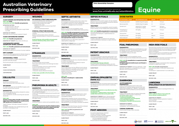
The equine Australian Veterinary Prescribing Guidelines poster. This document that outlines different antimicrobials for use in horses according to different diseases.
Funding for these guidelines was provided by the Australian Veterinary Association (AVA), Animal Medicines Australia (AMA) and AgriFutures Australia.
These guidelines would not have been possible without the considerable expertise and efforts of the Expert Panel: Associate Professor Laura Hardefeldt, Dr. Leanne Begg, Dr. Stephen Page, Professor Glenn Browning, and Professor Jacqueline Norris. We are also extremely grateful to the additional contributing authors.
The dedicated and skilled work of Project Manager Dr. Kellie Thomas is gratefully acknowledged, as are the contributions of the Project Steering Committee: Dr. Phillip McDonagh, Dr. John Messer, Professor James Gilkerson, and Dr. Melanie Latter. Open access publishing facilitated by The University of Melbourne, as part of the Wiley - The University of Melbourne agreement via the Council of Australian University Librarians.



Suggest a disease, syndrome or key evidence you think we should include.