
Traffic light - Horses
A document that outlines via a traffic light system, the different importance level of antimicrobials for use in horses.
The equine large intestine contains a large quantity of bacteria, amongst which anaerobes, Gram-positives and Gram-negative species. When Gram-negative bacteria die, endotoxin, a constituent of their cell wall, is released into the gut lumen. At normal rates of bacterial reproduction and death, only a relatively small amount of endotoxin is released. Absorption of this is mainly prevented by the mucosal barrier (epithelium, enzymes, IgA, and normal microflora) and the small amount that reaches the portal circulation is effectively removed by the liver. However, if there is disruption of the flora and/or the mucosal barrier, significant amounts of free endotoxin can be absorbed into the circulation, overloading the capacity of the liver to remove it. This results in clinical signs of endotoxaemia (see Section 7).
As most equine diarrhoea is hypersecretory, with loss of fluid and electrolytes into the gut lumen, dehydration occurs rapidly. Fluid losses in diarrhoea can be extremely high (up to 100 L per day), quickly resulting in hypovolaemia. In addition, hypovolaemia also develops due to endotoxaemic shock. Hypovolaemia and endotoxaemia both reduce tissue perfusion and oxygen delivery to the tissues, leading to hyperlactataemia. Common acid-base abnormalities include metabolic acidosis due to electrolyte losses (hyponatraemia) and hyperlactataemia, and metabolic alkalosis due to electrolyte (hypochloraemia) and protein losses. Protein is lost in the gastrointestinal tract via a protein-losing enteropathy. Progression of endotoxaemia is common, with a systemic inflammatory response syndrome (SIRS) deteriorating into multiple organ dysfunction syndrome (MODS), and eventually multi-organ failure (MOF) and death in severe or untreated cases. Laminitis, thrombophlebitis and acute kidney injury are common sequelae of MODS in patients with colitis. In severe and peracute cases, death can occur before the onset of passing of diarrhoea. Colic is common, especially in the early stages of the disease, before the onset of diarrhoea. Some cases develop neurological signs due to intestinal hyperammonaemia.
The aetiology of acute diarrhoea in adult horses in Australia includes infectious causes (Salmonella enterica, Clostridioides difficile and possibly Clostridium perfringens), carbohydrate overload, NSAID toxicity (right dorsal colitis), sand irritation, heavy infestations or rapid emergence of cyathostomins, dietary changes, and antimicrobial-associated diarrhoea. Equine proliferative enteropathy could also be a differential diagnosis in horses aged less than one year (see Chapter 5 in this section). Equine coronavirus is a recognised cause of acute diarrhoea and colic, sometimes in conjunction with neurological disease thought to be associated with hyperammonaemia (1), in the USA and Japan, but its significance in Australia is currently unknown. It has been detected in Australia in foals (Bailey, unpublished) and by PCR in the faeces of horses with colitis (2). Further investigation is required to evaluate the significance of a positive coronavirus PCR test in Australian horses with acute diarrhoea. Internationally, it can present as individual cases (2) but more commonly is associated with herd outbreaks (1).
The different aetiologies present very similarly, and it is impossible to distinguish them from each other without knowledge of the risk factors of the case and additional testing. The literature would suggest that in up to 50% of cases, a definitive diagnosis cannot be reached, despite extensive diagnostic testing.
Diarrhoea can occur following the use of antimicrobial drugs in adult horses, generally within the first week after initial administration. High-grain diets and the stress of training and performance are also thought to contribute. Oral antimicrobials, such as trimethoprim-sulphonamide and doxycycline have been implicated, but parenterally administered antimicrobials, such as cephalosporins and penicillin, can also result in diarrhoea. Oral administration of penicillins, macrolides (in adults), and oxytetracycline are strongly associated with antimicrobial-associated diarrhoea and these drugs should never be administered orally in adult horses. However, many antimicrobials used in the adult horse have the capacity to alter normal gut flora, regardless of route, and may result in colitis, so clients should be warned of the risks (3). The resulting diarrhoea can be life-threatening. Clostridioides difficile and Salmonella spp. have been equally implicated in antimicrobial-associated diarrhoea.
(see 8. Salmonellosis in this section)
Clostridial diarrhoea is caused by the Gram-positive, anaerobic, spore-forming bacteria. In adult horses, Clostridioides difficile is the most important species. The role of Clostridium perfringens remains unclear, although recent detection of novel toxins has renewed speculation about the importance of this pathogen (4). Other species have also been sporadically implicated, including C. septicum, C. cadaveris and C. sordellii. Previous administration of antimicrobial drugs is common. Any cause of stress, including hospitalisation, transportation and sudden dietary changes, can predispose to disease. Clostridial organisms are present in the soil and are a normal inhabitant of the gastrointestinal tract of horses. Disease associated with clostridia is mediated by enterotoxins – for C. difficile, 2 large molecular weight toxins, toxin A and toxin B, are shed early in the course of disease. Under conducive conditions, C. difficile spores germinate, and vegetative forms colonise the intestinal mucosa and produce toxins A and B. Horses may also be infected by direct ingestion of vegetative forms. Both toxins are believed to synergistically damage the mucosal epithelium by different mechanisms. In a 2023 study in Western Australia, C. difficile was detected in 38% of horses with gastrointestinal signs and 30% of horses without gastrointestinal signs, underscoring the lack of specificity of culture as a diagnostic tool in clinical cases. Only 55% of cases that carried C. difficile harboured a toxigenic strain in this study (5). C. perfringens enterotoxicosis is mediated by C. perfringens enterotoxin (CPE), alpha toxin (CPA), beta toxin (CPB), beta2 toxin (CPB2) or necrotising enterotoxin (NetF).
Cyathostomins are a group of small strongyle parasites. The larvae are pathogenic, but not the adults. Although the incidence of clinical disease associated with parasitism has decreased in recent decades with the availability of more effective anthelmintics, small strongyle infestations have been increasingly important due to the development of resistance to anthelmintics and the poor efficacy of current anthelmintics against the encysted larval stages. Cyathostomin infection occurs through the faecal-oral route. Larval stages travel to the caecum or colon, where they encyst in the intestinal mucosa and mature before re-emerging as adult worms. This re-emergence most commonly occurs in winter and early spring (6) and causes physical disruption of the intestinal wall, resulting in diarrhoea. The factors that contribute to the maturation and synchronous emergence of inhibited cyathostome larvae are poorly understood, but removal of adult worms from the intestinal lumen through ageing or anthelmintic treatments is thought to stimulate the inhibited stages to develop. Encysted third-stage larvae may persist in nodules (hypobiosis) for as long as two years. Synchronous larval emergence has been demonstrated to be associated with general dysbiosis of the intestinal flora (6).
After sudden access to or an increase in grain (i.e., carbohydrate) feeding, the hydrolytic and/or absorptive capacity of the small intestine is overwhelmed and a portion of the ingested carbohydrate passes into the cecum undigested, where it undergoes rapid fermentation, with increased production of lactate and gas, a decrease in caecal and colonic pH and an increase in endo- and exotoxin production, associated with increased intestinal permeability, favouring increased absorption of endotoxin. High risk changes to diet include sudden introduction to grain feeding or an abrupt increase in the amount of grain concentrate, feeding of large grain meals (even in horses adapted to such feeds), or grazing of lush pasture or first-cut forage with a high content of rapidly fermentable substrate, such as fructan and simple sugars (7).
Right dorsal colitis (RDC) has traditionally been thought to be associated with the oral administration of phenylbutazone, but there have also been sporadic cases associated with administration of flunixin meglumine or meloxicam, and a combination of NSAIDs may predispose to disease. The aetiology of RDC and the reason lesions are confined to the right dorsal colon are not fully understood. NSAIDS inhibit cyclooxygenase (COX), in the arachidonic acid pathway, reducing prostanoids, which convey some of the harmful effects of inflammation. However, these enzymes are also part of the physiological mechanisms for maintaining blood flow to the gastrointestinal mucosa and kidney. A lack of ability to restore mucosal barriers in the face of NSAID therapy was thought to cause RDC, but experimental models have failed to prove this pathogenesis. Further work is necessary to understand this disease. Also unknown is why some horses develop RDC while on treatment with NSAIDs and others do not, regardless of the drug, dose or duration of therapy, although many cases may only show very mild signs or be subclinically affected (8). Horses with inappetence and/or dehydration are thought to be more at risk, so extra care should be taken with these cases. Administration of phenylbutazone at standard doses (6 mg/kg/day) to healthy horses with concurrent 50% water restriction can induce disease in as little as 5 days (9). In clinical cases, gross lesions involve haemorrhage on the serosal surface, along with ulceration of the mucosa and oedema of the colon wall. Full necropsy examination often reveals lesions in other areas, including the oral cavity, stomach and kidneys (10).
Sand causes enteropathy by physical irritating the mucosa of the colon, resulting in colitis. Some horses actively eat sand, while others ingest soil and sand accidentally. Underfeeding or inadequate roughage or feeding from a sandy surface have been suggested to predispose to accidental eating of sand. Regardless, it is common for horses on sandy pastures to consume sand, although not all develop an enteropathy, and the reason for this individual variation is unknown. It is suggested that underlying colonic pathologies, such as inflammatory bowel disease, can lead to decreased clearance of sand in some cases. Clinical signs can include acute colic, recurrent colic, diarrhoea and unexplained weight loss.
History is very important when assessing colitis cases. Recent medical treatments, including anthelmintics, NSAIDS and antimicrobials, diet, and a history of any stressful events should be ascertained.
Clinical signs can range from mild to peracute, with sudden death. The classic triad of clinical signs are fever and diarrhoea along with leukopaenia. Haematology and biochemistry are critical to quantify dehydration, electrolyte loss, protein loss and organ dysfunction, as well as acid-base status and coagulation cascade activation. Moderate to severe haemoconcentration is common and may mask hypoproteinaemia and hypoalbuminaemia. Azotaemia is also very common, and it may be difficult to differentiate between renal and prerenal changes, as urine is generally difficult to obtain until fluid therapy is commenced. Monitoring of coagulation factors is valuable in cases where progression to disseminated intravascular coagulation is of concern, although testing is limited in many hospitals.
Lactate can be measured on a hand-held device. These devices are practical and financially viable for use in equine clinics and in ambulatory practice. Normal lactate in mature horses is < 0.7 mmol/L. Measurement can be used for diagnostic and prognostic information, as well as monitoring the response to therapy. In severe shock, lactate may transiently increase following initiation of intravenous fluid therapy due to release from tissues into the circulation with correction of perfusion.
Rectal examination may be useful in early cases to differentiate impending diarrhoea from other causes of colic, such as fluid faecal content passing over an impaction in the pelvic flexure.
Faecal samples should be submitted for PCR or culture for Salmonella spp. (see Chapter 8 in this section). PCR assays (not quantitative) for the A and B toxins of C. difficile and the genes for enterotoxin of C. perfringens are commercially available. However, the latter is of questionable value, as C. perfringens enterotoxin is of low virulence. A definitive diagnosis of clostridial diarrhoea can only be made when there is positive culture and demonstration of toxins. This is challenging as clostridial culture requires anaerobic techniques that are not widely available in private diagnostic laboratories, and the toxin test is not quantitative, so small amounts of toxin, possible from normal flora, can result in a positive toxin test.
Diagnosis of larval cyathostominosis is challenging, as clinical disease can occur before the parasites shed eggs or the larval stages in the faeces. Macroscopic examination of the faeces may be useful if there are large numbers of larvae present (redworms). A serum antibody test has recently been developed (11) that appears to be useful for detecting mucosal and luminal cyathostominosis, but this test is not yet available commercially.
Abdominal ultrasound is a very useful tool for evaluating the colon. Normal colonic wall thickness is 0.3-0.4 cm or less. All causes of colitis can result in thickening of the colonic wall, but thickening does not always occur. Thickening of the colonic wall is not pathognomonic for colitis, as it can also occur in other syndromes, such as colonic volvulus and inflammatory bowel disease. In horses with RDC, there is substantial thickening of specifically the right dorsal colonic wall, with a distinct hypoechoic layer (oedema) bordered by hyperechoic layers on both the serosal and mucosal sides, and the wall typically looks corrugated. A normal right ventral colon differentiates RDC from other causes of colitis, as the colon is generally diffusely thickened in other inflammatory or infectious causes of colitis. Sand is difficult to detect on ultrasound.
Abdominal radiographs are recommended if sand enteropathy is suspected.
Salmonella detection protocols are described in Chapter 8 of this section.
Horses with 2 of the 3 classical signs of colitis (fever, diarrhoea and leukopaenia) should be managed in isolation because of the risk of contagious and zoonotic diseases.
Referral of cases that have watery diarrhoea and signs of endotoxaemia (with or without fever), to a facility with 24 h care and isolation facilities is encouraged to provide intensive care and high-volume fluid therapy and improves the probability of a successful outcome.
The main treatment for horses with diarrhoea is symptomatic. Fluid therapy to correct dehydration, hypovolaemia and electrolyte loss is critical, and large volumes are usually required. Fluids can, in mild cases, be administered orally, but in moderate to severe cases oral fluid therapy is not sufficient or can even be contra-indicated (if ileus is present), and intravenous fluid therapy is essential. The water absorptive capacity of the colon (the section of the gastro-intestinal tract that absorbs water) is limited and compromised due to pathology and dysfunction of the colon and due to poor gastro-intestinal perfusion secondary to shock. Hartmann’s solution is preferred over normal saline (0.9% NaCl), as normal saline is acidifying, whereas Hartmann’s solution is alkalinising because it contains lactate, which is converted to bicarbonate in the liver. This is an advantage in colitis, where metabolic acidosis is the predominant acid-base abnormality. Hartmanns is also polyionic and therefore causes less electrolyte disturbances when given in large volumes than normal saline. Hypertonic saline (7% NaCl at 4 ml/kg IV) can lead to rapid volume expansion in severely hypovolaemic cases, but must be followed with isotonic fluids, either administered orally (via a nasogastric tube or providing free access to them) or intravenously. A resuscitation bolus with isotonic fluids of up to 60-80 ml/kg can be administered if needed. Ongoing fluid requirements can be estimated from maintenance needs, the degree of dehydration, and ongoing losses through the diarrhoea. Ongoing losses are difficult to estimate and may be large. Frequent monitoring of PCV, total protein, electrolytes and lactate are recommended to guide fluid therapy.
Treatment to address endotoxaemia is addressed elsewhere (see Section 7).
Protein loss is a common feature of acute colitis and can be significant and cause decreasing colloid oncotic pressure. Therefore, in the face of hypoproteinaemia, fluid therapy can easily lead to formation of oedema. Even though hydration is a vital treatment for colitis, it is also important to avoid overhydration and not to prolong use of fluid therapy longer than needed to minimize formation of oedema. Colloids, ideally plasma (up to 20 ml/kg) can be required. The cost of plasma for adult horses is substantial. Synthetic colloids are also available, with Hetastarch (10 ml/kg/day) the most widely used, although Pentastarch and Tetrastarch have also been administered to horses with colitis, hypoproteinaemia and low colloid oncotic pressure. Evidence for their use is weak, but in a retrospective study evaluating the use of Hetastarch, a smaller proportion of cases (47%) receiving Hetastarch survived than cases treated with plasma (80% survival), even though they had similar disease severity on admission (12). There has been concern about adverse effects associated with treatment with synthetic colloids, but none were recorded in the 23 horses that received Hetastarch alone in this study. Further studies are needed to evaluate the benefits of these products. At this time, in colitis cases with severe hypoproteinaemia, when plasma is not available or cost-prohibitive, synthetic colloids can be considered to improve colloidal oncotic pressure. Administration by constant rate infusion is recommended when using synthetic colloids to increase colloidal oncotic pressure (13).
Non-steroidal anti-inflammatory drugs are integral to address endotoxaemia, SIRS and pain. Flunixin meglumine remains the most used NSAID at either the so-called ‘anti-endotoxic dose’ (0.25 mg/kg IV q 8 h), which has no anti-inflammatory effects, or at a higher dose (0.5 – 1.1 mg/kg IV q 12 h) (See Section 7 for more information on managing endotoxaemia). The authors recommended higher doses of flunixin meglumine for combatting inflammation and pain in addition to endotoxaemia. However, NSAIDs should be avoided in cases with right dorsal colitis. If pain cannot be managed with NSAIDS (or without NSAIDs, in right dorsal colitis), use of other analgesic medications is recommended (opioids, alpha2 agonists, lidocaine constant rate infusion). In cyathostominiasis cases, steroid therapy (prednisolone or dexamethasone) can be indicated, instead of NSAIDs, to reduce the intestinal inflammation (6).
In general, antimicrobials are not indicated in cases of colitis. Antimicrobial use results in intestinal dysbiosis, which may prolong recovery and shedding of pathogens in colitis cases. In addition, there is no evidence of a more rapid recovery in cases of salmonellosis after antimicrobial therapy. Translocation of bacteria across the damaged intestinal mucosa has been demonstrated, but there is no evidence that antimicrobial therapy improves the prognosis. Historically, clinicians have treated horses with broad-spectrum prophylactic antimicrobials when neutropaenia was severe. However, there are many clinicians managing colitis cases with severe neutropaenia without antimicrobial therapy with no adverse effects. If secondary infections develop, then antimicrobial therapy should be targeted to these infections.
It is appropriate to avoid potentially nephrotoxic drugs as much as possible until onset of fluid therapy or until creatinine normalises.
Cryotherapy for all four feet may reduce the incidence of laminitis. Ideally, cryotherapy should be applied for the duration of the developmental phase of laminitis, so horses exhibiting endotoxaemia should be treated from the first clinical examination (see Section 7). There is an excellent review by van Eps, (14) of mechanism and recommendations for cryotherapy.
Re-instauration of a healthy flora, by administration of probiotics, is often suggested, but evidence for this is lacking (15). Faecal microbiota transplantation (administration of faecal liquid from a healthy horse to a sick horse) can be performed and has been shown to be safe. Significant advantages to patients are yet to be demonstrated (16) and optimal methods and timing of this treatment remain unclear, however this treatment is common in horses. Donors should be healthy, have not been exposed to antimicrobials in the past 6 months, and free from potential gastrointestinal pathogens, such as Salmonella spp. and gastrointestinal parasites. Ideally donors should be from the same farm and housed under the same conditions as the recipient. Recipients should have antimicrobial therapy discontinued prior to transplant and be pretreated with a protein pump inhibitor (omeprazole). Faeces are removed from the rectum of the donor horse and mixed with warm water or isotonic saline then the mixture blended and strained. A 2-3 L volume is administered by nasogastric tube for an average 450 kg horse. Free choice long stem early cut hay should be offered as soon as practical following faecal microbiota transplantation. The procedure should be repeated daily until there is improvement in faecal consistency, or for up to 3 days. A fresh transplant should be prepared daily (17).
Antimicrobial-associated diarrhoea: Antimicrobial therapy should be discontinued.
Clostridial diarrhoea: Metronidazole is indicated (15 mg/kg PO q 8 h or 25 mg/kg PO q 12 h) for 3 days. Resolution of diarrhoea is often rapid following metronidazole therapy. Resistance has not been documented in Australia. Treatment should be reserved for cases where clostridial infection has been documented with concurrent toxin gene detection. Because the action of the metronidazole is required within the gastrointestinal tract, oral metronidazole treatment is recommended. The intravenous and rectal routes have not been evaluated and transport into the gastrointestinal tract is unknown.
Cyathostominosis: The preferred treatment is with moxidectin (0.4 mg/kg PO). Fenbendazole (10 mg/kg PO q 24 h for 5 days) can also be used, but there is increasing resistance of strongyles to fenbendazole. Prednisolone at 1 mg/kg PO q 24 h for 24-48 h is recommended in clinically affected horses prior to and during administration of anthelmintics to control inflammation in the colon.
Right dorsal colitis: There are four principles of treatment: 1, avoid further NSAID use; 2, minimise stress; 3, dietary modification; and 4, targeted medication. Large-volume fibre should be eliminated from the diet to remove bulk from the colon and favour healing. Pelleted feed alone, or with small quantities of green grass, should be fed for 3-6 months. Oil can be used to increase calories in the diet, if necessary, and corn oil has been demonstrated to increase gastric PGE2 and promote mucosal healing. Misoprostol is a synthetic PGE1 analogue that can be administered at doses of 2-5 µg/kg every 6-12 h, but should be handled with caution, as exposure can cause abortion in people. Sucralfate and psyllium husks have also been used, but the efficacy of these treatments is unknown.
Sand enteropathy: Combination therapy with psyllium and MgSO4 (Epsom salts) (both at 1 g/kg) via nasogastric tube once daily for multiple days is the preferred method of treatment. Further, the collective findings of published research suggest that administering psyllium in feed as a preventative has no advantage over removing access to sand (18).
The prognosis is guarded. Referral to a hospital that can provide 24 h intensive care is recommended, as retrospective studies have reported mortality rates of between 19 and 42%.
Serial blood lactate measurement can be used to guide the prognosis. In retrospective studies, non-survivors had the highest lactate concentrations (mean of 5.35 mmol/L) at admission and after 72 h (mean of 2.35 mmol/L), compared to survivors (means of 1.75 mmol/L at admission and 0.75 mmol/L after 72 h) (19). Another study found 4-8 h lactate concentration, 24 h lactate concentration and the percentage reduction in lactate concentration (≥ 30% at 4-8 h and ≥ 50% at 24 h) were significantly associated with survival. No clear “cut-off value” for non-survival has been elucidated, emphasising the importance of serial measurements and examination of trends in lactate concentration, and full assessment of all clinical aspects of the patient, rather than any one value in assessing the prognosis.
The prognosis for right dorsal colitis and larval cyathostominosis may be worse than for other aetiologies, with mortality rates of approximately 60% reported. Antimicrobial-associated diarrhoea has been shown in multiple studies to have a significantly higher mortality than other causes of diarrhoea.
A document that outlines via a traffic light system, the different importance level of antimicrobials for use in horses.
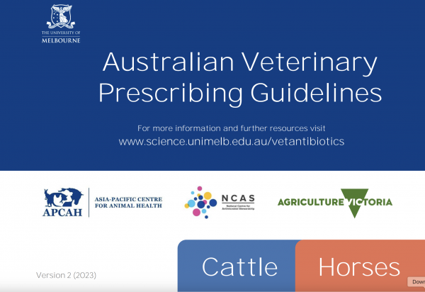
The Australian Veterinary Prescribing Guidelines cattle and horse flipbook, detailing antimicrobials for use in cattle and horses.
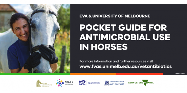
The equine Australian Veterinary Prescribing Guidelines for antimicrobial use as a pocket guide booklet.
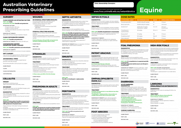
The equine Australian Veterinary Prescribing Guidelines poster. This document that outlines different antimicrobials for use in horses according to different diseases.
Funding for these guidelines was provided by the Australian Veterinary Association (AVA), Animal Medicines Australia (AMA) and AgriFutures Australia.
These guidelines would not have been possible without the considerable expertise and efforts of the Expert Panel: Associate Professor Laura Hardefeldt, Dr. Leanne Begg, Dr. Stephen Page, Professor Glenn Browning, and Professor Jacqueline Norris. We are also extremely grateful to the additional contributing authors.
The dedicated and skilled work of Project Manager Dr. Kellie Thomas is gratefully acknowledged, as are the contributions of the Project Steering Committee: Dr. Phillip McDonagh, Dr. John Messer, Professor James Gilkerson, and Dr. Melanie Latter. Open access publishing facilitated by The University of Melbourne, as part of the Wiley - The University of Melbourne agreement via the Council of Australian University Librarians.


Suggest a disease, syndrome or key evidence you think we should include.