
Traffic light - Horses
A document that outlines via a traffic light system, the different importance level of antimicrobials for use in horses.
Endometritis is a normal physiological process in the mare, enabling sperm, seminal plasma, bacteria and other debris that accumulates within the uterus during oestrus or breeding to be removed by a combination of both immunological and mechanical clearance mechanisms(5). Failure of this normal physiological process within the first 48 hours after breeding (6) is referred to as persistent breeding-induced endometritis or PBIE. Mares susceptible to PBIE have been identified as having a compromised immune system, in addition to weak mechanical defences, rendering them “susceptible” to uterine contamination and potential infection, which may lead to irreversible chronic degenerative changes in the uterus (Figure 15.1). Equine endometritis is one of the major causes of subfertility in the mare (7).

Figure 15.2. The aetiology of post breeding endometritis in the mare. Source: Morris LHA, McCue PM, Aurich C. Equine endometritis: a review of challenges and new approaches. Reproduction (Cambridge, England) 2020;160:R95-R110. (5)
Bacterial endometritis occurs when a mare fails to resolve the physiological endometritis and bacteria remain in the uterus and proliferate. The most common bacterial species isolated from the mare’s uterus are Streptococcus equi subspecies zooepidemicus (S. zooepidemicus) and Escherichia coli (8). However, a number of other species of bacteria and fungi have been isolated from the mare’s uterus (Table 15.1).
Table 15.1.
Microorganisms | Features | |
Bacteria | Streptococcus equi subspecies zooepidemicus | Gram positive, opportunistic bacteria, potentially venereally transmitted |
Escherichia coli | Gram negative, opportunistic, facultatively anaerobic | |
Pseudomonas aeruginosa | Gram negative, aerobic, potentially venereally transmitted | |
Klebsiella pneumoniae | Gram negative, opportunistic, facultatively anaerobic, potentially venereally transmitted | |
Staphylococcus spp. | Gram positive, opportunistic, facultatively anaerobic | |
Taylorella equigenitalis | Gram negative, venereal, microaerophilic, severe purulent endometritis. Not currently present in Australia. Notifiable disease. | |
Enterobacter cloacae | Gram negative, opportunistic, facultatively anaerobic | |
Proteus spp. | Gram negative, opportunistic, facultatively anaerobic | |
Fungi | Candida spp. | Yeast, causes 58-69% of fungal endometritis cases |
Aspergillus spp. | Mould with septate hyphae, causes 25-26% of fungal endometritis cases | |
Mucor spp. | Mould with aseptate hyphae, causes 5-12% of fungal endometritis cases |
(Adapted from Canisso IF, Segabinazzi LGTM, Fedorka CE. Persistent breeding-induced endometritis in mares — a multifaceted challenge: from clinical aspects to immunopathogenesis and pathobiology. International Journal of Molecular Sciences 2020;21:1432. (6))
Endometritis can be further complicated by the ability of many of these organisms to form a biofilm, with approximately 80% of isolates collected from the equine uterus capable of forming a biofilm (6). A biofilm is formed by the secretion of an exopolysaccharide matrix that allows microorganisms to evade the host’s immune system by creating a physical barrier resistant to penetration by both immune cells and antimicrobials (9). This effectively protects bacteria from traditional intrauterine antimicrobial therapy and, in fact, has promoted antimicrobial resistance in these species (10). By evading the host immune system, these organisms may lie dormant within the mare’s uterus without any clinical or cytological evidence of inflammation, delaying diagnosis and treatment (11, 12, 13).
Failure to identify and treat infectious endometritis may result in chronic degenerative changes to the uterine endometrium and the formation of endometrosis (14). These pathological changes can be identified histologically as severe endometrial fibrosis and cystic dilation of uterine glands (14, 15), and eventually result in infertility.
In the normal mare, the physiological endometritis following breeding is resolved within 48 h, with the successful clearance of debris, uterine fluid, seminal plasma and sperm. This presents a conundrum for the clinician, who ideally needs to identify mares that will have PBIE prior to breeding, so treatment can be initiated prior to closure of the cervix. The cervix increases in tone and effectively closes under the influence of rising progesterone post-ovulation. A history of fluid accumulation post-breeding is useful information, but there are also other diagnostic tests that can help to identify individuals with PBIE.
A careful reproductive tract examination of the broodmare, including a physical examination of the external reproductive tract and a trans-rectal ultrasonographic examination of the internal reproductive tract should be performed.
Mares susceptible to PBIE have compromised systemic and physical defence mechanisms. Often these mares will have poor external reproductive conformation, including breakdown of the three barriers to external contamination - vulvar conformation, the vestibulo-vaginal seal and the cervix (16). Age and parity results in acquired anatomical defects of the reproductive tract, with poor perineal and vulvar conformation, cervical incompetence and a pendulous uterus identified as significant structural issues in susceptible mares(5, 17). These anatomical defects can be identified during a reproductive tract examination of the broodmare. Surgical correction of the conformational defect should be addressed in these cases. A simple Caslick’s vulvoplasty may be the first surgical line of defence, followed by perineal body repair, urethral extension or uteropexy to correct more severe defects and to prevent further disease of the reproductive tract. It is important to note that a uteropexy should not be considered a last resort technique in mares. Performing this surgery earlier in the mare’s career may help to restore the normal position of the reproductive tract, thereby preventing both vaginitis and endometritis. Correction of these anatomical defects should improve the mare’s resistance against PBIE and, in turn, improve fertility and reduce the incidence of infectious endometritis.
Evidence of inflammation within the uterus can be identified as inappropriate oedema for the stage of the oestrus cycle and the presence of fluid within the uterine lumen. A mare can be diagnosed as susceptible when > 2 cm of fluid can be detected by trans-rectal ultrasonography during oestrus or within 36 hours of breeding, demonstrating a failure of both mechanical and immunological clearance mechanisms (5). This may be exacerbated by the position of the uterus over the pelvic brim, resulting in a dependant or pendulous uterus. In cases where uterine fluid accumulation is identified prior to breeding, an endometrial culture is recommended.
Once a presumptive diagnosis of PBIE is made, a breeding management plan can be formulated.
The significant difference between PBIE and infectious endometritis is the presence of a pathological organism within the uterine lumen. Often the conditions are not mutually exclusive, with untreated PBIE often progressing to infectious endometritis (5). Bacterial or fungal contamination of the uterus may occur at the time of mating, ascend from the external environment or via iatrogenic introduction. Infectious endometritis presents with similar clinical signs to PBIE, including intrauterine fluid accumulation and inappropriate uterine oedema for the stage of the oestrus cycle. A vulvar discharge may also be present and vaginoscopy may assist in determining the source of the discharge to differentiate between endometritis and vaginitis. Infectious endometritis caused by Taylorella equigenitalis is not currently present in Australia and thus will not be discussed in this section.
A definitive diagnosis of infectious endometritis can be made from an endometrial sample following culture and cytological examination. Endometrial samples for culture can be taken using a double guarded swab, low volume uterine lavage or uterine biopsy. Care should be taken to ensure that only the endometrium is sampled using any of these methods. Uterine sampling is inherently problematic as all sampling equipment passes through the potentially contaminated caudal reproductive tract. Therefore, careful cleaning and preparation of the vulva prior to vaginal entry is important to reduce false positives due to contamination and prevent iatrogenic infection. Whilst the double guarded swab technique is quick and easy to perform, low volume lavage has the advantage of sampling the entire surface of endometrium resulting in greater sensitivity (18, 19). The technique for performing a low volume uterine lavage for endometrial culture is described by Dr Michelle LeBlanc and a link can be found in the further reading section of this chapter (20). Uterine biopsy is not performed routinely for culture, but may be considered, particularly if the clinician suspects that there may be bacteria dormant within the crypts of the endometrium or biofilm-producing organisms (13).
Interpretation of results should be based on consideration of the findings of the breeding evaluation of the mare. If there is evidence of uterine fluid and excessive oedema in addition to a cloudy flush, positive culture and evidence of inflammation on cytology, then the treatment plan is usually straight forward. Culture and cytology should be performed together and interpreted based on the clinical presentation. When a uterine lavage has been performed to obtain the sample, characterisation of the efflux fluid and presence of debris can also help to rule out false positive culture results (20). Small number of single bacterial colonies are likely to be the result of contamination, especially in the absence of any additional supportive information. A confident diagnosis of bacterial endometritis can be made when there is evidence of inflammation (2-5 polymorphonuclear cells per microscope field at 400 x) and/or a pure bacterial culture (21). However, E. coli infections tend to suppress inflammation and organisms within a biofilm or in a latent state, such as Streptococcus spp., may not stimulate recruitment of immune cells into the uterine lumen. Activation of such organisms with a commercial growth medium, such as B-Activate (22), or disruption of the biofilm with a suitable biofilm disrupter (23, 24) may assist detection of these organisms and ensure adequate treatment to clear the infection. It is also important to note that the failure to culture bacteria when there is evidence of inflammation is not necessarily a false negative result. In these instances, care should be taken to look for clinical evidence of pneumovagina and urine pooling and consider the relevant breeding history of the mare.
Treatment of endometritis focuses on the physical removal of fluid, bacteria and debris from the uterus, in addition to antimicrobial therapy when a positive culture result has been obtained. Targeted treatment of the uterus is preferred over systemic antimicrobial therapy, which does not specifically target just the problematic organisms, is more likely to promote antimicrobial resistance, changes the microflora of the gastrointestinal tract and contributes to environmental contamination. Treating the uterus also allows the dose and duration of antimicrobial treatment to be reduced and refined. Systemic antimicrobial use should only be considered in cases of chronic endometritis or when infusion into the uterus is contraindicated.
The uterus is an easily accessible cavity allowing uterine lavage to be performed safely to reduce contamination and allow better penetration of antimicrobials. Uterine lavage and ecbolic (oxytocin) therapy are the mainstays of treatment of endometritis. Sterile saline or lactated Ringer’s/Hartmann’s solution are suitable for uterine lavage and should be followed by administration of oxytocin at 10-20 IU IM or IV to further promote uterine contractility. Following appropriate cleaning and preparation of the vulva, a gloved (sterile, where possible) hand is passed through the labia and into the vagina. A sterile Foley catheter is passed through the cervix and the cuff inflated cranial to the internal cervical os. Sterile fluid can then be instilled into the uterine lumen and syphoned back out. This step is repeated until the efflux is clear. In mares with a dependant uterus and fluid accumulation, this step may need to be repeated twice daily and oxytocin administered as frequently as every 2 h.
Post-breeding uterine lavage can be considered in mares with known PBIE and should be performed at 4 hours after breeding to allow sufficient time for spermatozoa involved in fertilisation to move through the utero-tubal papillae into the oviducts. The normal physiological immune response of the uterus occurs as soon as 30 minutes after breeding with a peak at around 4-6 h (25). Uterine lavage at this stage helps to mitigate the inflammatory response in mares with PBIE by physically removing the antigenic products of breeding and promoting uterine contractility. If uterine lavage is unable to be performed, oxytocin should be administered to these mares in this same time frame to promote uterine contractility and clearance. However, this clearance is far more effective when combined with uterine lavage.
Historically, intrauterine treatment of mares with prophylactic antimicrobials was encouraged to improve pregnancy rates in thoroughbred mares. However, more recent publications have contradicted this recommendation, demonstrating that prophylactic infusion of antimicrobials does not improve pregnancy rates in mares and, furthermore, can contribute to antimicrobial resistance (26, 27). Whilst there has been an increased reliance on post-breeding therapy, including the use of intrauterine antimicrobials, over the past 20 years, the incidence of pregnancy loss and live foaling rates have remained unchanged (28). If there is no evidence of a pathogen, based on cytology and culture, treatment using intrauterine antimicrobials cannot be recommended. Prophylactic therapy should therefore include uterine lavage and oxytocin, which have been shown to not only improve pregnancy rates (27), but also mitigate the effects of PBIE in susceptible mares.
Antimicrobials should only be considered when a pathogen has been cultured from the uterus. Often it may take several days to receive culture and susceptibility test results. During this time uterine lavage and treatment with oxytocin can be performed as pretreatment prior to antimicrobial administration. This effectively reduces the bacterial overgrowth, thereby reducing the reliance on antimicrobials to clear the infection. Furthermore, the presence of debris or excessive fluid in the uterus may inhibit the action of infused antimicrobials or excessively dilute them to subtherapeutic concentrations (29). A biofilm reducer may also be considered as part of the treatment regimen at this stage to eliminate any potential biofilms, thereby improving penetration of antimicrobials once the susceptibility test results are available.
Biofilm reducers or mucolytics have been recommended by several researchers and veterinarians. However, it is important to note that not all biofilm reducers are effective for all bacterial species (Table 15.2). Furthermore, occasionally the introduction of a biofilm reducer into the uterus may result in an inflammatory effect (e.g. H2O2), so care should be taken when selecting the timing of infusion. In suspect cases of infectious endometritis without confirmatory culture results, infusion of a biofilm reducer or mucolytic may result in biofilm breakdown, releasing the pathogen and enabling it to be cultured.
Table 15.2. Compounds used to degrade biofilms.
Bacteria | Degradation of biofilm mass | Killing of bacteria within a biofilm |
E. coli | Tris-EDTA/Tricide Acetylcysteine H2O2 Dimethylsulphoxide (DMSO) | Acetylcysteine H2O2 |
Streptococcus zooepidemicus | Tris-EDTA/Tricide H2O2 Dimethylsulphoxide (DMSO) Hypochlorous acid | Tris-EDTA/Tricide H2O2 Dimethylsulphoxide (DMSO) Hypochlorous acid |
Pseudomonas aeruginosa | Tris-EDTA/Tricide H2O2 | Acetylcysteine |
Klebsiella pneumonia | H2O2 |
(Adapted from: Ferris RA, McCue PM, Borlee GI, Loncar KD, Hennet ML, Borlee BR. In Vitro Efficacy of Nonantibiotic Treatments on Biofilm Disruption of Gram-Negative Pathogens and an In Vivo Model of Infectious Endometritis Utilizing Isolates from the Equine Uterus. J Clin Microbiol. 2016;54(3):631-9. (30))
Other mucolytics such as CeragynÔ are not currently available in Australia but also show promising results for the removal of biofilm from pathogenic equine reproductive bacteria.
Several different antimicrobials have been infused into the uterus to eliminate bacteria or fungi causing endometritis. However, the dose rates of many of these antimicrobials have limited supporting data to establish an MIC for specific organisms. Intrauterine infusions should be administered following appropriate cleaning and preparation of the vulva and perineum. Clean equipment should be used to ensure that no further contamination occurs at the time of infusion that may compromise the effectiveness of the antimicrobial.
The volume of solution instilled into the uterus should be sufficient to achieve uniform distribution of the antimicrobial over the luminal surface without excessive losses from reflux. Therefore, volumes of 20-60 mL are usually recommended. Antimicrobials can be diluted in sterile saline, water for injection or, in some cases, 7.5% sodium bicarbonate to buffer and dilute to the infusion volume.
Fungal endometritis is difficult to treat, with recurrent infections common because of the anatomical defects that contribute to the problem. Organisms are often not detected because long incubation times are needed to culture them. In addition, getting a definitive speciation is also difficult. More commonly, fungi are detected by observation of them in cytological samples, further highlighting the importance of performing both cytological examination of samples and culture. Mares with fungal endometritis usually have a history of bacterial endometritis, with intrauterine antibacterial therapy predisposing them to fungal infection (29). Effective treatment of intrauterine fungal infections requires a combination of uterine lavage, uterine disinfection and intrauterine antimicrobials (Table 15.3). The aim of uterine lavage and disinfection is to physically remove all fungal elements from the uterus, and create a hostile environment to inhibit further growth. Antiseptic solutions that have been advocated for this purpose include 1% hydrogen peroxide solution, 2% acetic acid solution (vinegar), 0.1 - 0.2% povidone iodine solution and 20% dimethylsulphoxide solution (29, 31). These antiseptic solutions are diluted in 1 L of 0.9% saline solution to achieve the desired concentration and daily lavage is performed during oestrus (5 - 7 days). Following uterine antiseptic lavage, a repeat culture should be performed, as lavage is sufficient treatment in some cases. In recalcitrant fungal endometritis, further treatment using intrauterine antifungals is required. It is important that any anatomical defects are addressed surgically to prevent further infection. If immunocompromise of the individual is suspected, then investigation of endocrine disorders, such as pituitary pars intermedia dysfunction or equine metabolic syndrome, is warranted.
In addition to these therapies, treatment with immunomodulators has been investigated as an adjunctive therapeutic approach. Whilst non-steroidal anti-inflammatory drugs and glucocorticoids have been used successfully to reduce inflammation following breeding, there are some drawbacks to their administration, including impairment of the normal reproductive hormonal cascade. Bacterial extracts, such as Mycobacterium phlei cell wall extract (Settle), have been used to enhance the innate humoral immune response by decreasing pro-inflammatory cytokines and increasing anti-inflammatory cytokine production (6). Mares treated with this product had reduced immune cell infiltration into the uterus, mimicking the immune response of a resistant mare. This immune stimulant can be administered to the broodmare as part of the routine breeding management of the mare to normalise the immune response to breeding (32).
Drug | IU infusion dose | Comments |
Ampicillin | 2 g | Soluble product, high dilution to prevent irritation |
Ceftiofur sodium | 1 g | *Reserved for resistant organisms, use only with culture and susceptibility results |
Gentamicin sulphate | 1 g | Buffered with equal volumes of 7.5% sodium bicarbonate or a large volume (60 mL) of sterile saline |
Penicillin (procaine) | 3 – 6 g | High dilution (60 mL) to prevent irritation |
Enrofloxacin | 250 mg | High dilution (60 mL) to prevent irritation *Caution: may cause severe, acute ulcerative endometritis and possible fibrosis. Ciprofloxacin (600 mg) may be used as an alternative when indicated by culture and susceptibility testing |
Clotrimazole | 400 – 700 mg | Intrauterine infusion q 24 h for 7 days, moulds and yeasts |
Miconazole | 500 - 700 mg | Intrauterine infusion q 24 h for 7 days, moulds and yeasts |
Nystatin | 0.5 - 2.5 x 10⁶ IU | Intrauterine infusion q 24 h for 7 days, yeasts only |
Amphotericin B | 100 - 200 mg | Intrauterine infusion q 24 h for 7 days, moulds and yeasts |
Fluconazole | 100 mg | Intrauterine infusion q 24 h for 7 days, yeasts only |
A document that outlines via a traffic light system, the different importance level of antimicrobials for use in horses.
The Australian Veterinary Prescribing Guidelines cattle and horse flipbook, detailing antimicrobials for use in cattle and horses.
The equine Australian Veterinary Prescribing Guidelines for antimicrobial use as a pocket guide booklet.
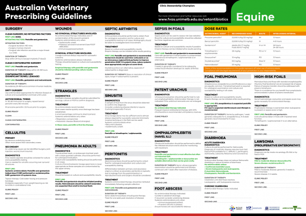
The equine Australian Veterinary Prescribing Guidelines poster. This document that outlines different antimicrobials for use in horses according to different diseases.
Suggest a disease, syndrome or key evidence you think we should include.