
Traffic light - Horses
A document that outlines via a traffic light system, the different importance level of antimicrobials for use in horses.
Oesophageal ulceration occurs when gastric emptying is delayed because of pyloric obstruction.
Equine Squamous Gastric Disease (ESGD) occurs in the squamous mucosa, predominantly along the margo plicatus. Primary ESGD occurs in an otherwise healthy gastrointestinal tract and is the most common form of this disease. Secondary ESGD is due to delayed gastric outflow, as a sequela of other diseases, such as pyloric stenosis, severe EGGD or inflammatory bowel disease (IBD).
Horses evolved as grazing animals and secrete hydrochloric acid (HCl) continuously in the stomach, so, if they are stabled and only fed intermittently, acid concentrations increase in the stomach, damaging the cranial squamous mucosa. Fibre is critical to the pathogenesis and likely acts in two protective ways: firstly, by increasing the saliva produced by chewing, which has a buffering effect on stomach acid; and secondly, through the formation of ‘roughage balls’ in the stomach to limit acid splashing. Fibre size is therefore an important factor, with long fibre length particularly important. Crib-biting and other stereotypical behaviours are also associated with ESGD.
The caudal glandular part of the stomach secretes mucus and bicarbonate, providing protection from the normal acidic environment. Ulceration in this area, Equine Glandular Gastric Disease (EGGD), is less well understood, but NSAID therapy blocking prostaglandin production, gastritis, or an altered microbiota may be involved. Training and environmental stress are also thought to play a part in glandular ulceration, but diet has not been implicated.
ESGD is very common, with prevalences of 80 - 90% in horses in training and 30-60% in horses not in training and in wild populations. The prevalence of EGGD is more variable, with rates of between 15 and 65% reported.
Helicobacter pylori has not been cultured from the stomach of horses and is not thought to be involved in the pathogenesis of gastric ulcers in horses (30).
Reported clinical signs vary and are often non-specific. They include colic, weight loss or poor body condition, poor coat condition, reduced appetite, diarrhoea, bruxism, a Flehmen response, behavioural changes and poor performance.
Gastroscopy using a 3 m endoscope, after 12 h of fasting, is the only reliable ante-mortem method of definitely diagnosing gastric ulceration (31). The oesophagus is best visualised when withdrawing the scope from the stomach, when the oesophagus is dilated between peristaltic waves. Mucosal biopsies are of limited value in detecting underlying disease compared to full-thickness biopsies. For EGGD, assessment of the clinical relevance of ulceration should not be based on the endoscopic appearance alone. Instead, the relevance of a lesion should be assessed in the light of clinical signs and the response to treatment. Faecal occult blood testing is unreliable.
ESGD can be graded based on its visual appearance on endoscopy. Grading allows lesions to be monitored.
For EGGD, the recommendation at this time remains not to assign a grade to these lesions, but rather describe the lesions by anatomical location, distribution, severity and appearance (31).
Proton-pump inhibitors are currently the best acid suppressive therapy available and there is sufficient evidence for their use for ESGD. Growing evidence also supports the use of acid suppression for EGGD, along with mucosal protectants.
Enteric-coated omeprazole is recommended (1-2 mg/kg PO q 24 h). Administration should be in the morning, approximately 30 min prior to feeding after fasting overnight. The long-acting injectable formulation of omeprazole appears to be more successful for treating EGGD when administered at 5-day intervals, although it was not registered in Australia at the time of writing these guidelines and local injections site reactions can occur. Treatment is generally for 3 weeks for ESGD and 3-4 weeks for EGGD.
Sucralfate (12 mg/kg PO q 12 h) is recommended in combination with omeprazole for EGGD (31), although doses of up to 20-30 mg/kg q 6-8 h are commonly used. Sucralfate should be administered at least 30 min after omeprazole.
Misoprostol has also been proposed as a treatment for EGGD, but further evidence is needed before a recommendation can be made.
Management changes are very important for ESGD. Ad libitum roughage, or at least 2% of bodyweight per day of good-quality roughage, should be consumed. Afternoon exercising may also reduce acid exposure and limiting exercise to 40 minutes per day may be beneficial. For EGGD, duration of exercise is less important, but numbers of days of exercise have been associated with disease, so it is recommended that horses get 2 or, ideally, 3 full days rest per week (32). In addition, reducing stress may help in reducing EGGD, although evidence is lacking.
Oesophageal inflammation due to choke is treated by removing the food obstruction. Feeding a soft diet usually results in resolution of oesophageal inflammation. Sucralfate may be useful in coating ulceration.
The prognosis is good for oesophageal inflammation once the obstruction is resolved.
Healing rates for ESGD are good with oral omeprazole monotherapy (67-100%), but less successful for EGGD (14-25%).
A document that outlines via a traffic light system, the different importance level of antimicrobials for use in horses.
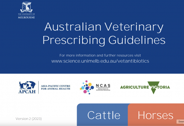
The Australian Veterinary Prescribing Guidelines cattle and horse flipbook, detailing antimicrobials for use in cattle and horses.
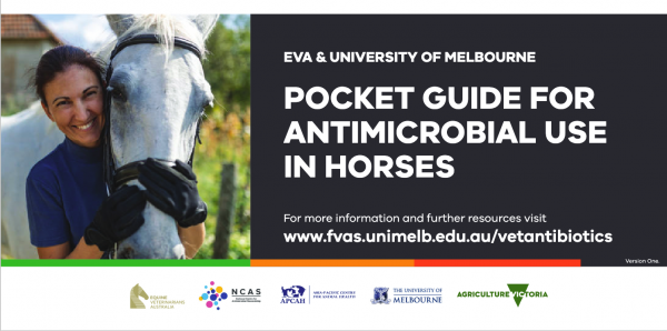
The equine Australian Veterinary Prescribing Guidelines for antimicrobial use as a pocket guide booklet.
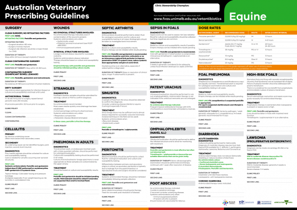
The equine Australian Veterinary Prescribing Guidelines poster. This document that outlines different antimicrobials for use in horses according to different diseases.
Funding for these guidelines was provided by the Australian Veterinary Association (AVA), Animal Medicines Australia (AMA) and AgriFutures Australia.
These guidelines would not have been possible without the considerable expertise and efforts of the Expert Panel: Associate Professor Laura Hardefeldt, Dr. Leanne Begg, Dr. Stephen Page, Professor Glenn Browning, and Professor Jacqueline Norris. We are also extremely grateful to the additional contributing authors.
The dedicated and skilled work of Project Manager Dr. Kellie Thomas is gratefully acknowledged, as are the contributions of the Project Steering Committee: Dr. Phillip McDonagh, Dr. John Messer, Professor James Gilkerson, and Dr. Melanie Latter. Open access publishing facilitated by The University of Melbourne, as part of the Wiley - The University of Melbourne agreement via the Council of Australian University Librarians.


Suggest a disease, syndrome or key evidence you think we should include.