
Traffic light - Horses
A document that outlines via a traffic light system, the different importance level of antimicrobials for use in horses.
This page will outline antimicrobial prescribing gruidelines for the following musculoskeletal conditions:
Click here (https://doi.org//10.1111/avj.70003) to view the guidelines in their entirety.
Clostridial myonecrosis is a syndrome of severe necrotizing soft tissue infection associated with Clostridium spp. It typically presents as peracute emphysematous soft tissue swelling in the region of an injection or wound within hours of the inciting cause. Intramuscular injection of several common drugs, including flunixin meglumine, ivermectin, omeprazole, antihistamines, phenylbutazone, dipyrone, vitamin B complex and synthetic prostaglandins, has been implicated. Disease following injection of flunixin meglumine appears over-represented and the association with wounds seems less common. Central to the aetiology is the inoculation of the organisms and the creation of anerobic foci in which the organisms can proliferate, sometimes facilitated by irritant medications. Clostridium perfringens and C. septicum are the species that have most often been reported as causative organisms, but other Clostridium species have also been implicated. Rapid identification of cases is critical, as prompt treatment can improve survival rates. Horses can develop clinical signs and die within 48 - 72 h.
Horses are generally febrile and have moderate to severe depression and inappetence. They are generally stiff and reluctant to move. Severe toxaemia and cardiovascular collapse can occur soon after clinical signs develop, with diffuse intravascular coagulation and acute renal failure as consequences.
Soft tissue swelling, with palpable subcutaneous emphysema, is very common, especially when disease affects the cervical region, and this generally leads to a presumptive diagnosis. When the disease affects the gluteal region, subcutaneous emphysema may be a less reliable clinical sign. The overlying skin may initially feel hot, but quickly becomes cold, tough and insensitive due to the underlying necrosis. Aspiration of fluid from the affected region, or upon myotomy/fasciotomy, reveals large parallel-sided, Gram-positive rods. Ultrasonography can be useful to identify gas shadows within deeper soft tissues and pockets of necrotic tissue to target for fasciotomy.
All horses that develop soft-tissue swellings acutely after IM, SC or inadvertent perivascular administration of drugs and have systemic signs of illness should be assessed by examining a Gram-stained smear of an aspirate from the affected area. Release of exotoxins by the bacteria causes muscle necrosis and destruction of white blood cells. Absorption of the exotoxins into the circulation causes widespread damage to the endothelium, liver and muscles, leading quickly to death.
An aggressive approach to therapy is prudent when clostridial myonecrosis is suspected or confirmed. Medical and surgical therapy should be pursued.
Medical:
Surgical:
Fenestration of the emphysematous area is indicated to improve oxygenation, reduce swelling and facilitate debridement of necrotic tissue. This is typically performed standing with only light systemic sedation. Due to the extensive necrosis, only light local analgesia is needed. Multiple vertical incisions, approximately 2.5 cm apart, should be made through the muscles in the affected area. Exposed tissues can be irrigated and many different solutions have been advocated (hydrogen peroxide, chlorhexidine, crystalline penicillin), but there is no evidence that any one agent is more beneficial. Daily hydrotherapy should follow to keep the area clean and facilitate granulation. Owners should be warned of the significant soft tissue and skin sloughing that will probably occur over the medium to long term.
In a series of 37 cases, 27 horses (73%) survived to discharge, with a median duration of hospitalisation of 12 days, although treatment was necessary following discharge in all cases. There has been speculation that survival rates may differ depending on the Clostridium species involved, but there is insufficient evidence to make such distinctions.
Immune mediated myositis is a rare, but severe, disease that typically causes rapid and severe symmetrical wasting of the topline muscles, often following exposure to, or vaccination against, Streptococcus equi subspecies equi (strangles). The disease appears to predominately affect Quarter horses and related breeds and has been associated with a specific genetic variant in these breeds. Atrophy may progress to involve 50% of the horse’s muscle mass within 1 week and may lead to generalised weakness. The reason specific muscle groups are affected is unclear.
Muscle loss is caused by inflammatory destruction of fast-twitch muscle fibres.
Haematological abnormalities are generally minor, with relatively mild increases in CK and AST, given the severity of disease.
Although clinical signs are suggestive, a muscle biopsy is required to confirm the diagnosis.
Horses with concurrent evidence of strangles should be treated with antimicrobials (penicillin) and it is prudent to avoid IM injections. Administration of corticosteroids seems to immediately improve clinical signs and prevent further progression of muscle atrophy. Dexamethasone is generally administered (0.1 mg/kg IV for 3 days) and then the dose tapered over 1 month. Some recommend switching to prednisolone (not prednisone) for oral administration (1 mg/kg) following the initial 3 days of therapy.
Full muscle mass can be regained within weeks to months, but recurrence of atrophic episodes is common (~40% of horses). Euthanasia because of severe muscle loss and poor quality of life is common.
Synovial structures can become infected by direct inoculation following a wound, by iatrogenic introduction with intra-synovial injections or by haematogenous spread. Although haematogenous spread is more common in foals, it can occur in adult horses. Early diagnosis and aggressive treatment with lavage and appropriate antimicrobial therapy is critically important, as chronic disease is associated with higher mortality. The most common bacterial isolates from wounds are Enterobacter spp., Staphylococcus spp., Streptococcus spp. and Pseudomonas spp., but infections are often mixed and can involve anaerobic bacteria. Iatrogenic infections are often caused by Staphylococcus spp.. Studies of the efficacy of addition of prophylactic antimicrobials (such as amikacin) to intra-articular medications (such as corticosteroids) have not demonstrated a reduction in joint sepsis, so this prophylactic use is not recommended. Aseptic joint preparation and adequate injection technique are sufficient to prevent sepsis (3). Polysulphated glycosaminoglycans (PSGAGs) are the only exception to this, with the current recommendation being to use concurrent intra-articular antimicrobials to reduce the risk of infection (4).
Although the use of intra-articular antimicrobials for treatment of joint sepsis is widespread, there are important clinical knowledge gaps about the appropriate intra-articular doses to use, the potential for contribution to the development of antimicrobial resistance with overuse, and whether there are long-term adverse side effects of the cytotoxicity of local antimicrobial treatment for cartilage. The intraarticular administration of amikacin can induce dose-dependent increases in cartilage degradation products and biomarkers of inflammation. It is thought that concurrent administration of other medications, such as hyaluronic acid, may mitigate antimicrobial-induced cytotoxicity (5).
Intravenous regional limb perfusions (IVRLP) are used to treat distal limb infections by isolating the limb from the systemic circulation with a tourniquet and infusing a high concentration of an antimicrobial intravenously. The antimicrobial diffuses passively into all tissues throughout the isolated region until the tourniquet is removed, after approximately 20 min. Ceftiofur sodium administered at a dose of 2 g diluted in 60 mL of sterile saline as an IVRLP maintained regional plasma concentrations above MIC (1 µg/mL) for 12 h, and subcutaneous tissue concentrations above MIC for 24 h, but bone concentrations were only above MIC until immediately after tourniquet removal (6). Inflamed joints have been shown to have increased concentrations of amikacin after IVRLP, when a 5 mg/kg dose (one-third of the systemic dose) was diluted in 60 mL of sterile saline, and the concentrations reached the recommended Cmax-to-MIC ratio of 8 (MIC of 16 µg/mL) in most inflamed joints, but not in normal joints (7). There are many published reports describing variations in this technique, including the dose used, the dose volume and the concentration of perfusate, the dosing interval, the type, method and duration of tourniquet application (with or without an Esmarch bandage) and whether the technique is performed standing or under general anaesthesia, so the optimal method for performing IVRLP has not been established and the apparent clinical benefits of the technique are often difficult to confirm and quantify (8). For detailed recommendations on intra-synovial and intra-venous regional perfusion, see the next section (Section 12).
Horses with septic synovial structures are generally febrile and non-weight-bearing lame in the affected leg, with effusion into the infected synovial structure. Synovial fluid analysis reveals an elevated nucleated cell count (> 2.5 x 10⁹/L), with a neutrophilia (> 80%) and an elevated protein concentration (> 25 g/L). Synovial fluid should be submitted for culture and susceptibility testing.
Fair to good with early detection and prompt treatment of infection. In recent studies 84% and 90% survived to discharge (mean duration of antimicrobial therapy is 12 days), but only 54% and 65% returned to function (9, 10).
A document that outlines via a traffic light system, the different importance level of antimicrobials for use in horses.
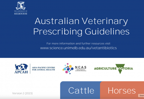
The Australian Veterinary Prescribing Guidelines cattle and horse flipbook, detailing antimicrobials for use in cattle and horses.
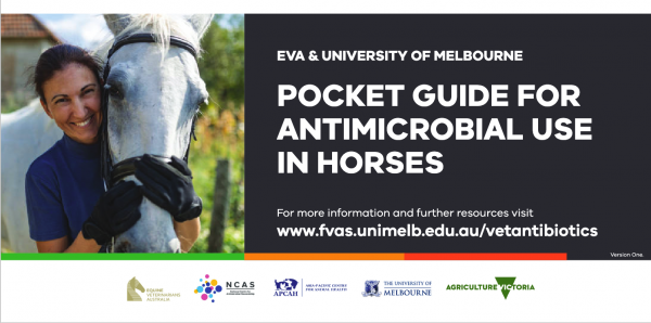
The equine Australian Veterinary Prescribing Guidelines for antimicrobial use as a pocket guide booklet.
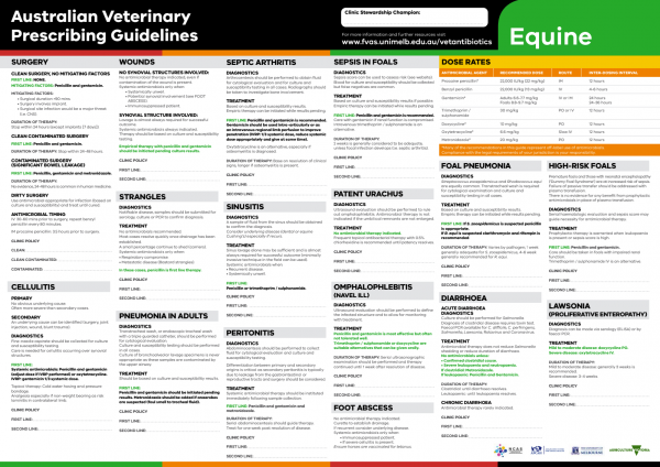
The equine Australian Veterinary Prescribing Guidelines poster. This document that outlines different antimicrobials for use in horses according to different diseases.
Funding for these guidelines was provided by the Australian Veterinary Association (AVA), Animal Medicines Australia (AMA) and AgriFutures Australia.
These guidelines would not have been possible without the considerable expertise and efforts of the Expert Panel: Associate Professor Laura Hardefeldt, Dr. Leanne Begg, Dr. Stephen Page, Professor Glenn Browning, and Professor Jacqueline Norris. We are also extremely grateful to the additional contributing authors.
The dedicated and skilled work of Project Manager Dr. Kellie Thomas is gratefully acknowledged, as are the contributions of the Project Steering Committee: Dr. Phillip McDonagh, Dr. John Messer, Professor James Gilkerson, and Dr. Melanie Latter. Open access publishing facilitated by The University of Melbourne, as part of the Wiley - The University of Melbourne agreement via the Council of Australian University Librarians.
![]()

Suggest a disease, syndrome or key evidence you think we should include.