
Traffic light - Horses
A document that outlines via a traffic light system, the different importance level of antimicrobials for use in horses.
Click here (https://doi.org//10.1111/avj.70003) to view the guidelines in their entirety.

Meningitis is inflammation of the meninges of the brain and spinal cord. The aetiology may be infectious or non-infectious. In horses, meningitis typically has an infectious aetiology. Organisms can invade the central nervous system (CNS) via traumatic injury, ascending infection, haematogenous spread (particularly in septic foals) or iatrogenic routes. Ascending infections can originate from the eyes, oral cavity, nasal passages, sinuses or osteomyelitis of the cranial bones or vertebrae. Inflammation of the meninges may lead to increased permeability of the blood-brain barrier, vasculitis, and central nervous system oedema.
Bacterial meningitis and meningoencephalitis are uncommon diseases of horses. They generally have high case fatality rates. Horses are particularly difficult to treat because of their large size and the challenge and hazards associated with nursing care.
Neurological deficits are common, with changes to mentation, ataxia, recumbency or other gait abnormalities seen in most cases. Cranial nerve deficits are also very commonly detected, with head tilt (cranial nerve VIII), nystagmus (cranial nerve VIII) and strabismus (multiple cranial nerves) most common, followed by an absent menace response (cranial nerve VII), absence of pupillary light response (cranial nerve II and/or III) and facial nerve paralysis (cranial nerve VII).
Fever, tachycardia and tachypnoea may or may not be present. Haematological analysis commonly reveals changes in leukocytes (either leukocytosis or leukopaenia). Cerebrospinal fluid (CSF) sample collection, from the site closest to the lesion (usually the atlanto-occipital cistern), is essential for making a diagnosis and CSF is generally grossly abnormal, with a cloudy or aserosanguinous appearance most common. Cytology of CSF reveals moderate to marked suppurative inflammation. Intra or extracellular bacteria are only seen in a third of cases. Culture should be pursued, but is frequently unsuccessful, even in cases where bacteria are seen on cytology. Many different pathogens have been isolated from cases, reflecting the wide range of predisposing causes.
Imaging should be undertaken in cases of suspected trauma. Computed tomography (CT) or magnetic resonance imaging may be necessary to detect fractures but are not widely available.
Intravenous fluid support, anti-inflammatory drugs, sedatives and intensive nursing care are of critical importance.
Broad spectrum antimicrobials are indicated and, although the blood-brain barrier is probably disrupted, antimicrobials that penetrate the blood-brain barrier should be prioritised. Trimethoprim/sulphadiazine or oxytetracycline are good empirical choices. Penicillin, gentamicin and ceftiofur have poor penetration.
There is no information available to guide the duration of treatment because of the poor clinical outcomes in most cases.
Poor to grave, even with aggressive treatment. There are few clinical reports of successful treatment.
Otitis media refers to inflammation of the middle ear or tympanic cavity, which contains the three small ossicles. Otitis interna results in inflammation of the inner ear and bony labyrinth, where the cochlear (auditory) and vestibular nerves are located. The pathogenesis is unknown, but haematogenous spread of bacteria, ascending infection from the respiratory tract, extension of otitis externa or extension of guttural pouch infection may be responsible for disease.
Lesions in the area of the petrous part of the temporal bone, tympanic bulla and hyoid apparatus can result in clinical signs of otitis media/interna in horses.
Inflammation may result in osseous proliferation and thickening of the temporohyoid joint, resulting in fusion – known as temporohyoid osteoarthropathy (THO). It is unknown whether THO is a primary osteoarthropathy or occurs secondary to inner ear infection. Many, including these authors, do not believe that otitis media/interna is a frequent underlying pathology in THO.
Clinical signs of otitis media/interna result in deficits attributable to the facial and vestibulocochlear nerves (cranial nerves VII and VIII) and can include a head tilt, nystagmus, falling, circling, ataxia (worsened with blindfolding), muzzle deviation, decreased lacrimation, ear paresis, corneal ulceration, loss of the blink reflex and depression. The head tilt is towards the side of the lesion and animals tend to circle towards the lesion or lie on the side of the lesion.
Diagnosis is made based on clinical signs, radiographic findings, endoscopic examination of the guttural pouch, computed tomography and the results of tympanocentesis. Haematology is generally unremarkable, and horses are rarely febrile.
Radiographs reveal enlargement of the proximal portion of the stylohyoid bone and sclerosis of the petrous part of the temporal bone. Endoscopic examination may be more sensitive than radiography, as enlargement of the proximal stylohyoid bone can be observed.
Tympanocentesis is technically difficult because of the long ear canal of the horse. General anaesthesia is required and may be difficult to justify in an ataxic horse. Without tympanocentesis, only a diagnosis of THO can be made. However, this procedure is not undertaken in most cases and thus in a diagnosis of THO otitis media/interna is often presumed to be the primary disease.
Although clinical signs are generally unilateral, bilateral disease is common.
Surgical management of temporohyoid osteoarthropathy has been shown to improve survival compared to medical treatment only and there are a range of techniques now reported in the literature. Surgery is aimed at reducing the load on the temporohyoid articulation with the goal of reducing pain and preventing fracture or refracture of the petrous temporal bone. The surgical procedures are specialised and should be performed by suitably trained specialists.
In cases where bacterial otitis media/interna is suspected or confirmed, antimicrobial therapy with agents with high volumes of distribution should be selected to maximise penetration. Trimethoprim/sulphadiazine and chloramphenicol are examples of lipophilic antimicrobials with high volumes of distribution. Given the potential human complications with chloramphenicol exposure, trimethoprim/suphadiazine is the most frequently used antimicrobial.
There is no evidence to support a specific duration of treatment, but 7 days is a reasonable first course.
Guarded. In a retrospective study of 33 medically treated cases, most horses had residual cranial nerve deficits and maximal improvement took one year or longer. In a study of 24 surgically managed cases, nearly 90% substantially improved within one year, with most improvement occurring within six months, but only 50% of cases returned to athletic performance.
Vertebral body osteomyelitis is a rare but life-threatening condition in horses. The disease is thought to result from haematogenous spread of a pathogen in most cases, although trauma with subsequent bony infection can also occur. Foals are most commonly affected, but disease has been reported in adult horses, likely with a primary immunocompromising disease. The cervical and lumbar regions of the spinal column are the regions most commonly affected.
A number of different pathogens have been reported, but Rhodococcus equi appears to be over-represented in foals. Other pathogens include E. coli, Salmonella spp., Actinobacillus spp., Streptococcus spp. and others.
Clinical signs include:
Radiographic signs include:
Radiographic changes may not be seen until 2 - 8 weeks after the onset of clinical signs. Advanced diagnostic imaging with computed tomography is useful, but is not available in most areas of Australia. Thoracic radiographs should also be taken in 2 - 6 month old foals, as radiographic evidence of pneumonia would support a diagnosis of R. equi osteomyelitis, but cases have been reported in the absence of lung lesions (see more on R. equi in Chapter 4).
Haematological changes are consistent with inflammation, but are not specific for the disease.
Isolation of the organism in blood or urine can be attempted in cases where treatment is being pursued. Fine needle aspirates of the affected area can be submitted for cytology and culture.
Medical therapy alone is unlikely to be successful. If attempted, blood and urine cultures should be used to guide therapy when positive. A sample of fluid obtained by fine needle aspiration of the affected area can also be submitted for culture. Where empirical antimicrobial therapy is necessary, selection should be appropriate for R. equi in foals in the at-risk age group (clarithromycin at 7.5 mg/kg PO q 12 h PLUS rifampicin at 5 mg/kg PO q 12 h). It is important to note that this combination does not have any Gram-negative coverage and the addition of gentamicin is probably warranted.
In cases where R. equi is not suspected, broad-spectrum therapy with penicillin (procaine penicillin at 22,000 IU/kg IM q 12 h or benzyl penicillin at 12-16 mg/kg IV q 6 h) and gentamicin (6.6 mg/kg IV q 24 h for animals aged > 2 weeks) is suitable.
Surgical debridement of the lesion is probably necessary, but increases the risk of fracture or collapse of the affected vertebrae. Stabilisation of the area is probably necessary and requires specialist surgical knowledge. Culture of the debrided material should guide antimicrobial therapy.
Nursing care is critical and typically needs to be intensive.
Duration of therapy is unknown, but several weeks of therapy is likely necessary.
Grave. Successful treatment of one foal has been reported with an aggressive surgical technique.
A document that outlines via a traffic light system, the different importance level of antimicrobials for use in horses.
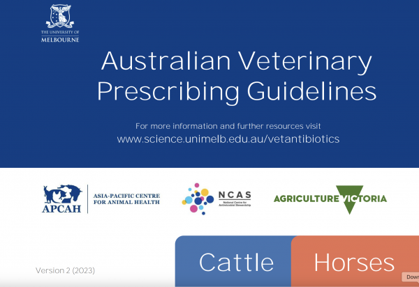
The Australian Veterinary Prescribing Guidelines cattle and horse flipbook, detailing antimicrobials for use in cattle and horses.
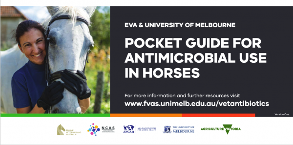
The equine Australian Veterinary Prescribing Guidelines for antimicrobial use as a pocket guide booklet.
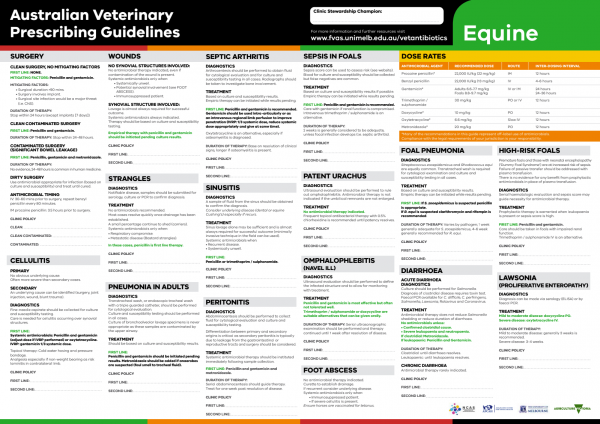
The equine Australian Veterinary Prescribing Guidelines poster. This document that outlines different antimicrobials for use in horses according to different diseases.
Funding for these guidelines was provided by the Australian Veterinary Association (AVA), Animal Medicines Australia (AMA) and AgriFutures Australia.
These guidelines would not have been possible without the considerable expertise and efforts of the Expert Panel: Associate Professor Laura Hardefeldt, Dr. Leanne Begg, Dr. Stephen Page, Professor Glenn Browning, and Professor Jacqueline Norris. We are also extremely grateful to the additional contributing authors.
The dedicated and skilled work of Project Manager Dr. Kellie Thomas is gratefully acknowledged, as are the contributions of the Project Steering Committee: Dr. Phillip McDonagh, Dr. John Messer, Professor James Gilkerson, and Dr. Melanie Latter. Open access publishing facilitated by The University of Melbourne, as part of the Wiley - The University of Melbourne agreement via the Council of Australian University Librarians.
![]()

Suggest a disease, syndrome or key evidence you think we should include.